Special Somatic Afferent Pathways
The Visual Pathways
Visual information is conveyed from the retina by the optic nerve. After entering the cranial cavity through the optic foramen, the nerve converges to meet its fellow in the optic chiasm on the ventral surface of the brain.
At the chiasm, there is a partial decussation of axons from each retina. The proportion of axons crossing to the opposite side is inversely correlated with the degree of binocular vision in a particular species. In ungulates, the binocular field of vision is small, and a very large percentage (85% to 90%) of fibers cross. Carnivores have a more binocular vision and a corresponding smaller proportion (75%) of axons cross in those species. Approximately 50% cross in primates, in which binocular vision is best developed. In birds, all axons were thought to cross, and it was considered that birds had no binocular vision—that is, that each eye was thought to see completely separate parts of the visual field. However, newer information indicates that some birds have an even larger field of binocular vision than humans.After reassortment of axons at the chiasm, these axons then continue on as the optic tracts, which arch over the lateral surface of the thalamus (Fig. 8.22/20). Most axons terminate within the lateral geniculate nucleus of the thalamus, which forms a swelling on dorsolateral surface of the thalamus. The axons of the second-order neurons then project, via the optic radiation within the internal capsule, to the visual cortex, which is located within the occipital lobe of the cerebrum. The visual cortex is the cortical region responsible for conscious visual perception (Fig. 8.45/6).
Those optic tract fibers that do not synapse in the lateral geniculate nuclei project onto various mesencephalic nuclei. A proportion of these optic tract axons synapse in the pretectal region, an area near the border between the thalamus and the midbrain.
Second-order neurons in the pretectal region then project to and synapse on neurons of the oculomotor nerve located in the midbrain that are responsible for reducing pupillary diameter in response to light. Optic tract fibers also synapse in the rostral colliculi, an important visual integration center in the midbrain responsible for controlling direction of gaze. Axons arising from the rostral colliculi also end on lower motor neurons in the cervical spinal cord and constitute the tectospinal tract, part of the so-called extrapyramidal motor system.Vestibular Pathways
The vestibular axons arising from the vestibular sensory apparatus in the inner ear travel within the common vestibulocochlear nerve (CN VIII) that enters the brain at the level of the trapezoid body. These axons then terminate on, or detach collateral branches to, neurons of the vestibular nuclei (Fig. 8.46/2). Some vestibular axons enter the caudal cerebellar peduncle to synapse within the vestibular portion of the cerebellum. The second-order axons that arise from neurons in the vestibular nuclei are divided between those that also pass to the cerebellum and the remainder, which travel to the spinal cord via the vestibulospinal tract and to the brainstem via the medial longitudinal fasciculus. Within the spinal cord, vestibulospinal axons project via a series of interneurons onto lower motor neurons of the ipsilateral ventral gray matter. The vestibulospinal tracts are part of the extrapyramidal system. Those second-order fibers that follow the medial longitudinal fasciculus (Fig. 8.46/4) and the reticular formation proceed to synapse on the nuclei of the cranial nerves supplying the external ocular muscles.
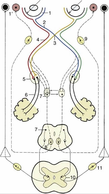
FIG. 8.45 A simplified schema of the visual and pupillary reflex pathways. Thick solid lines indicate special somatic visual fibers; thin solid lines, sympathetic fibers; and interrupted lines, parasympathetic fibers.
1, Retina; 1', dilated and constricted pupils; 2, optic nerve; 3, optic chiasm; 4, optic tract; 5, lateral geniculate nucleus; 6, optic radiation; 7, rostral colliculus and pretectal nuclei; 8, oculomotor nucleus (parasympathetic part); 9, ciliary ganglion; 10, lateral visceral efferent column; 11, cranial cervical ganglion.Finally, the axons that lead to conscious perception of vestibular stimuli travel from the vestibular nuclei via the lateral lemniscus and thalamic nuclei to a particular region of the cerebral cortex of the temporal lobe.
Auditory Pathways
The axons of the cochlear component of the vestibulocochlear nerve synapse within the dorsal and ventral cochlear nuclei located on the dorsal surface of the brainstem (Fig. 8.47/1 and 2). The second-order axons from the ventral cochlear nucleus then proceed to synapse within either an ipsilateral or a contralateral nucleus of the trapezoid body (Fig. 8.47/3). The pathway is then continued by axons of third-stage neurons carried within the lateral lemniscus. A proportion of these axons synapse within the nucleus of the lateral lemniscus (Fig. 8.47/5), a second group of axons travels to and synapses within the caudal colliculus (Fig. 8.47/6), and a third set of axons, concerned with the conscious perception of sound, travels farther rostrally to synapse in the medial geniculate nucleus of the thalamus, which in turn sends axons to the auditory cortex, located within the temporal lobe.
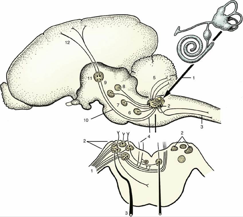
FIG. 8.46 A simplified scheme of the vestibular pathways. 1, Vestibular fibers in vestibulocochlear nerve; 2, vestibular nuclei; 3, vestibulospinal tract; 4, medial longitudinal fasciculus; 5, vestibulocerebellar tract; 6, abducent nucleus; 7, trochlear nucleus; 8, oculomotor nucleus; 9, red nucleus; 10, vestibulothalamic tract (in lateral lemniscus); 11, thalamic nuclei; 12, thalamocortical projection fibers.
The second-order axons that emerge from the dorsal cochlear nuclei join the ipsilateral or contralateral lateral lemniscus and thereafter follow the same courses as those that proceed from the ventral cochlear nuclei.