The basically similar structures enclosing the distal phalanx appear strikingly different.
Their origins as local modifications of skin are reflected in their retention of epidermal, dermal, and subcutis layers (though perhaps in greatly altered form). Nails, claws, and hoofs serve primarily to protect the underlying tissues, but each is also used for other purposes, such as scratching and digging or as a weapon.
The equine hoof, the most complex, reduces concussion on foot impact. Fig. 10.18 shows the correspondences among these appendages, each of which presents three parts: wall, sole, and associated pad. It is only in ungulates that the last forms part of the horny structure; it corresponds with the digital bulb of primates and the digital pad of carnivores.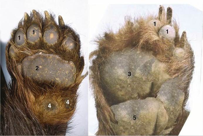
FIG. 10.14 Footpads of a bear, forelimb (left) and hindlimb (right). 1, Digital pads; 2, metacarpal pad; 3, metatarsal pad; 4, carpal pads; 5, tarsal pad, fused with the metatarsal pad.
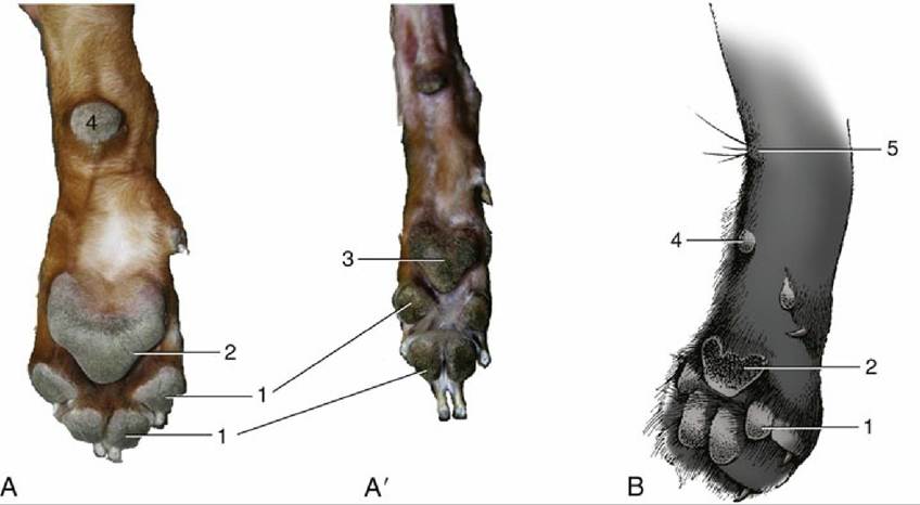
FIG. 10.15 Footpads of canine (A) forelimbs and (A') hindlimbs and of (B) feline forelimb. 1, Digital pads; 2, metacarpal pad; 3, metatarsal pad; 4, carpal pad; 5, carpal gland and associated tactile hairs.
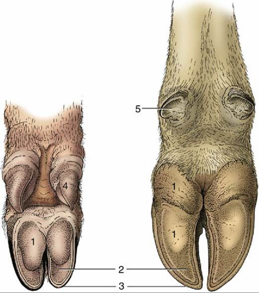
FIG. 10.16 Palmar surface of foot of the pig (left) and of a cow (right). 1, Bulb (digital pad) of hoof; 2, sole of hoof; 3, wall of hoof; 4, hoof of accessory digit; 5, rudimentary hoof of dewclaw.
The nail (wall) of primates grows from the epidermis covering a curved fold of dermis at its base. The epidermis under most of the nail produces a little horn that helps maintain adhesion as the nail gradually slides distally. The dermis under this rather unproductive portion of the epidermis is gathered into a few low, longitudinal folds (laminae) that interdigitate with corresponding epidermal laminae; increased dermoepidermal contact strengthens the bond between the nail and the deeper tissues.
The epidermis underlying the free border of the nail produces small amounts of soft "sole horn" (Fig. 10.18/2).The wall of the claw of carnivores can be likened to a nail that has been laterally compressed and so has obtained a sharp dorsal border. Its proximal part and the germinal layer from which it is derived are similarly shaped and are lodged with the associated dermis within the unguicular crest of the distinctively shaped distal phalanx (Fig. 10.18D). The epidermis deep to the wall is minimally productive. The dermis that covers the unguicular process fuses with the periosteum, and as with the primate nail, longitudinal interdigitations between dermal and epidermal laminae strongly bond the claw to the dorsal border of the bone. The space between the free margins of the wall on the undersurface of the unguicular process is filled with flaky "sole horn" (Fig. 10.18/5).
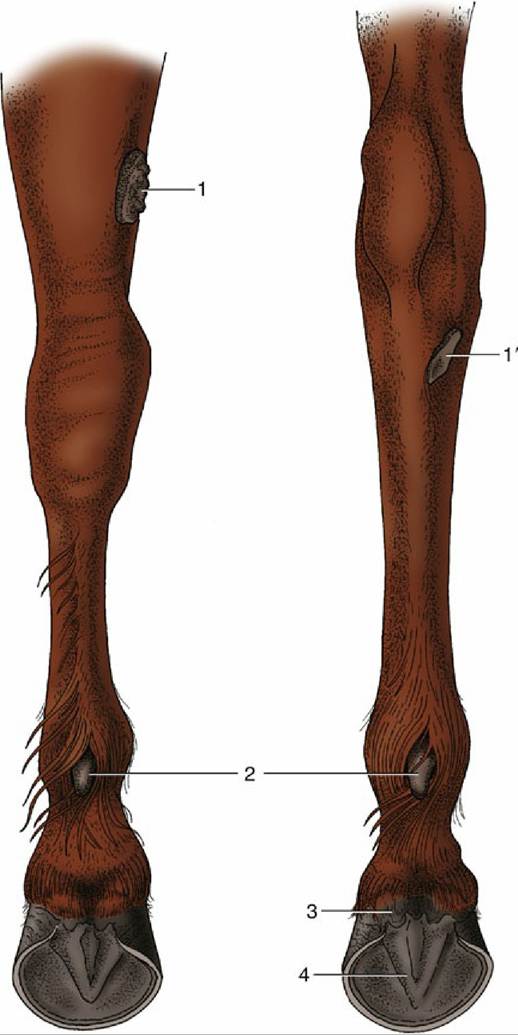
FIG. 10.17 Left forelimb (left) and left hindlimb (right) of the horse, caudal view. 1 and 1', Chestnuts above carpus and below hock, respectively; 2, ergots; 3, bulbs of the heels; 4, frog.
The wall of the horse's hoof is also strongly curved, and the sides are sharply inflected to form the so-called bars (Fig. 10.19E/2 "). The space between the bars is occupied by the frog, the part of the footpad that makes contact with the ground. The sole horn that fills the ground surface between wall and frog meets the wall at a junction known as the white line (zona alba; Fig. 10.19/5). The wall grows distally from the epidermis over a bulging (coronary) dermis* studded with numerous papillae directed toward the ground. The epidermis covering these papillae produces horn tubules that run distally, toward the weight-bearing margin of the wall. The tubules are embedded in less structured intertubular horn formed by the epidermis over the interpapillary regions of the dermis; the combination of horn types gives the tissue a finely striated appearance.
The (laminar) epidermis deep to the wall is again only minimally productive. It is arranged as several hundred well-formed laminae that tightly interdigitate with an equal number of dermal laminae (see Chapter 23, p. 600),bonding the wall to the underlying distal phalanx. One should remember that this is a living bond that allows the wall to slide gradually toward the ground, where its distal border is worn away. A band of soft horn (periople) lies over the external surface of the wall near its junction with the skin (Fig. 10.20/1). It descends with the wall and dries to a protective glossy layer. The band widens at the back of the hoof, where it covers the bulbs of the heels and part of the frog.
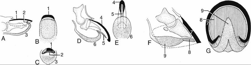
FIG. 10.18 Schematic representations of (A)-(C) nail, (D) and (E) claw, and (F) and (G) hoof. (A) Longitudinal section, (B) palmar surface, and (C) head-on view of human fingertip. (D) Longitudinal section and (E) palmar surface of canine claw. (F) Longitudinal section and (G) ground surface of equine hoof. 1, Nail (wall); 2, “sole horn” of nail; 3, bulb of finger; 4, wall of claw; 5, “sole” of claw; 6, digital pad; 7, wall of hoof; 8, sole of hoof; 9, frog.
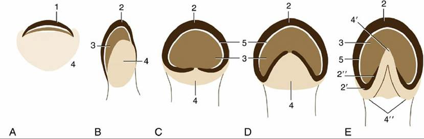
FIG. 10.19 An interpretation of the phylogenetic “development” of the horn structures associated with the distal phalanx. (A) Human fingertip. (B) Pig hoof. (C) Rhinoceros hoof. (D) Tapir hoof. (E) Horse hoof. 1,
Nail; 2, wall of hoof; 2' and 2", heel and bar (of horse); 3, sole; 4, footpad (bulb in human finger and pig); 4' and 4", frog and bulbs of the heels (of horse); 5, white line.
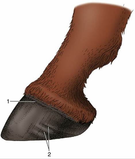
FIG. 10.20 Equine hoof. 1, Periople; 2, rings indicating uneven horn growth.
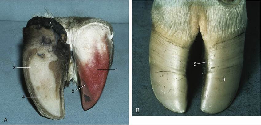
FIG.
10.21 (A) Bovine foot, palmar view. (B) Bovine foot, dorsal view. The horn shoe (epidermis) has been pulled off one digit in (A), exposing the dermis. 1, Dermis of bulb; 2, dermis of sole; 3, horn of bulb;4, horn of sole; 5, dorsal border of hoof; 6, abaxial surface of hoof.
The hoofs of ruminants and the pig, although resembling those of the horse in principle, differ in several respects: the wall is sharply bent to form a dorsal border (like that of the claw); the footpad (bulb) is relatively large and furnishes the entire caudal part of the hoof (Fig. 10.19B/4); the sole between the bulb and wall is small; and the interdigitating laminae are less developed (Fig. 10.21/2).
In all species, periods of disturbed or lessened horn production create ridges on the wall parallel to the formative region at the junction with the skin (Fig. 10.20/2).
Fuller accounts of these specializations are found in the appropriate later chapters.