» The Bladder and Urethra
The bladder is intra-abdominal in the young calf. In the adult, the bladder is in the pelvic cavity when empty but extends over the abdominal floor when distended. The neck within the pelvis is without a peritoneal covering and is attached to the pelvic floor by fat and loose connective tissue (see Figs.
29.7 and 29.8). Urine escaping from a ruptured bladder—a relatively common mishap, especially in steers—may infiltrate this tissue or enter the peritoneal cavity according to the site of the tear. There are the usual lateral and median ligaments.The relations of the bladder naturally vary. In the cow, it is always in contact with the cranial part of the vagina and the cervix and often with the body and horns of the uterus. Within the abdomen it makes contact with the dorsocaudal blind sac of the rumen and with the intestines (Fig. 29.11).
The urethra is much narrower than that of the mare and runs below the vagina, to which it becomes increasingly attached as it proceeds caudally. It opens into the vestibule through a median slit that is shared with the suburethral diverticulum (Fig. 29.11/13), a blind pouch extending cranially that is large enough to admit the end joint of a finger. The pouch can be a nuisance when catheterization is attempted. The urethralis muscle only covers the caudal part of the urethra, which more cranially is anchored to the floor by a short but strong ligament. The cranial fascicules of the urethralis muscle insert on a dorsal raphe that completes the encirclement of the urethra; the more caudal ones form a "U" shape that attaches to each side of the vagina and vestibule, enclosing both the diverticulum and the urethra.
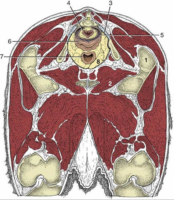
FIG. 29.8 Transverse section of the bovine pelvis at the level of the first caudal vertebra (cranial surface). The section passes through the obturator foramina. Note that the peritoneum covers only the dorsal surface of the vagina; the lateral and ventral surfaces are retroperitoneal at this level. (See Fig.
29.11 for the level of this section.) 1, Greater trochanter; 2, obturator foramen; 3, sacrosciatic ligament; 4, rectum; 5, rectogenital pouch; 6, vagina; 7, neck of bladder; 8, retroperitoneal fat.
The blood supply to these organs comes from the umbilical and vaginal arteries.