» The Rectum and Anus
Although the origin of the rectum is arbitrarily defined, its most caudal part is distinguished from the colon by a wider caliber and more muscular wall. The interior, marked by impermanent transverse folds, is generally distended with feces (Fig.
29.6).The colic mesentery continues as the mesorectum, which abruptly shortens to a mere 3 cm, before gradually decreasing further until it eventually disappears (Figs. 29.7 and 29.8), which brings the rectum into broad contact with the pelvic roof. In this process more and more of the rectal circumference becomes denuded of serosa until the last part is completely embedded in fat, which provides the cushion that allows the gut to adjust to changing circumstances. The close connection with the pelvic roof and walls is a handicap to rectal explorations, and for many purposes the hand must be carried forward into the more mobile colon (Fig. 29.9) (p. 707).
The anal canal is embraced by the pelvic diaphragm; the postdiaphragmatic part forms a low eminence presenting a short transverse slit through which the skin continues to provide the last stretch of the canal with a cutaneous epithelial covering. The anus is guarded by the usual two sphincters, and the striated external one exchanges fascicules with other muscles of the perineum (Fig. 29.10).
Most of the rectum is supplied from the cranial rectal artery, a branch of the caudal mesenteric, but the terminal section and the anal region are supplied by twigs from the caudal rectal artery, an indirect branch of the vaginal artery. The venous drainage is divided between the portal and systemic systems.
FIG. 29.7
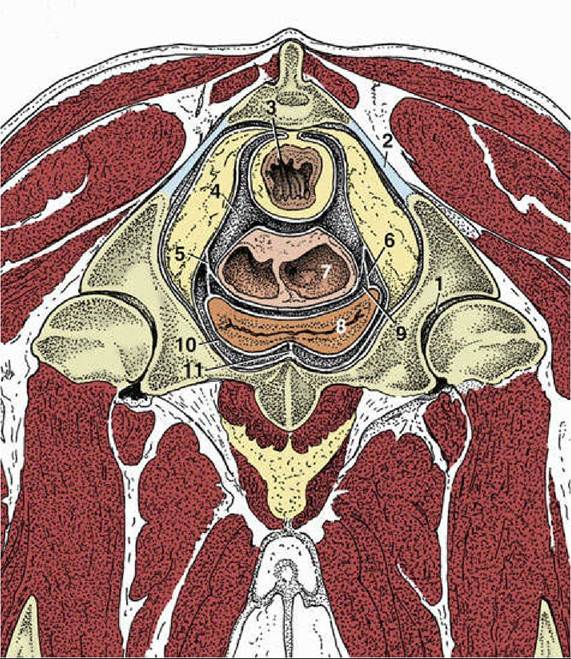
Transverse section of the bovine pelvis at the level of the hip joint (cranial surface). Note the
large amount of retroperitoneal fat in the pelvis. (See Fig. 29.11 for the level of this section.) 1, Hip joint; 2, sacrosciatic ligament; 3, rectum; 4, rectogenital pouch; 5, broad ligament of uterus; 6, lateral ligament of
bladder; 7, uterus sectioned where the two horns are conjoined; 8, bladder; 9, vesicogenital pouch; 10, pubovesical pouch; 11, median ligament of bladder.
More on the topic » The Rectum and Anus:
- The large intestine comprises the cecum, colon, rectum, and anal canal. In the carnivores such as the dog and cat, the colon is relatively small and the cecum is only a vestigial component.
- ANORECTAL DISEASES
- CONSTIPATION
- FECAL INCONTINENCE
- Anatomy
- Obstructive Intestinal Diseases
- DIAGNOSTIC CONSIDERATIONS
- Diarrhea in Neonatal Foals
- Abdominal Distention and Constipation
- Equine Herpesvirus Myeloencephalopathy