THE BLOOD VESSELS AND LYMPHATIC STRUCTURES OF THE FORELIMB
The axillary artery, the main supply of the limb, enters the axillary space after crossing the cranial border of the first rib, where it may be punctured (p. 539). It descends on the medial aspect of the arm in company
with the median and ulnar nerves and shortly becomes known as the brachial artery.
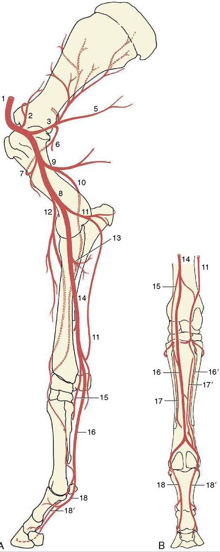
The trunk releases several branches to the muscles of the shoulder and arm, the most prominent being the subscapular artery, which follows the caudal border of the scapula, and the deep brachial, which disappears between the heads of theFigure 23-39 The major arteries of the right forelimb. A, Medial view. B, Palmar view. 1, Axillary a.; 2, suprascapular a.; 3, subscapular a.; 5, thoracodorsal a.; 6, 7, caudal and cranial circumflex humeral aa.; 8, brachial a.; 9, deep brachial a.; 10, collateral radial a.; 11, collateral ulnar a.; 12, transverse cubital a.; 13, common interosseous a.; 14, median a.; 15, radial a.; 16, 16', medial and lateral palmar aa.; 17, 17’, medial and lateral palmar metacarpal aa.; 18,18', medial and lateral digital aa.
triceps (see Figure 23-8). Just proximal to the elbow joint, lesser cranial and caudal branches (transverse cubital and collateral ulnar arteries, respectively) are detached for the muscles in the forearm; their further courses and connections are shown in Figure 2339/11,12. The brachial artery crosses the elbow cranial to the medial collateral ligament, where it can be palpated and the pulse evaluated, through the pectoralis transversus (Figure 23-40/5). Together with the median nerve it dips under the flexor carpi radialis caudal to the radius and soon gives off the common interosseous artery, which passes through the interosseous space to reach the craniolateral muscles of the forearm.
The main trunk, now redesignated the median artery (Figure 23-41/12), gradually works its way to the caudal surface of the forearm before dividing into three above the carpus.
The lesser branch (palmar branches of the median and radial artery) contribute the small palmar metacarpal arteries that accompany the interosseus muscle, while the main trunk passes through the carpal canal with the digital flexor tendons (Figure 23-15, B/19). It continues with these in the cannon where it becomes the medial palmar artery, the main artery to the digit and hoof. This inclines axially before splitting into the medial and lateral digital arteries above the fetlock. The digital arteries pass over the abaxial surfaces of the sesamoid bones (where they are palpable) and continue into the digit on each side of the flexor tendons; the lateral artery is reinforced by the small metacarpal arteries that join above the sesamoid bone (Figure 23-39/18'). The branches of the digital arteries distal to the fetlock are symmetrical. Dorsal and palmar branches are given off opposite PI, and these supply adjacent structures while forming a circle about the bone. A branch to the digital cushion is detached level with the pastern joint before the digital artery disappears by passing deep to the hoof cartilage. Dorsal and palmar branches detached opposite the middle of PII comport themselves similarly to the branches about PI but also take part in the supply of the dermis of the hoof. The dorsal and palmar terminal branches (to PIII) have been described (pp. 602 and 612); the palmar branches anastomose to form a terminal arch within the bone.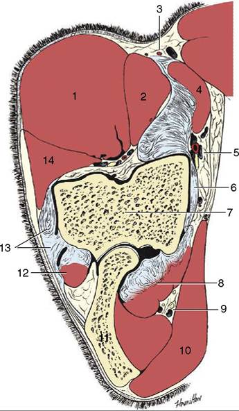
Figure 23-40 Transverse section of the left elbow. 1, Extensor carpi radialis; 2, brachialis; 3, medial cutaneous antebrachial nerve and cephalic vein lying on lacertus fibrosus; 4, biceps; 5, brachial vessels and median nerve; 6, medial collateral ligament; 7, humerus; 8, flexors arising from medial epicondyle of humerus; 9, ulnar nerve and collateral ulnar vessels; 10, tensor fasciae antebrachii; 11, olecranon; 12, ulnaris lateralis; 13, lateral collateral ligament; 14, common digital extensor.
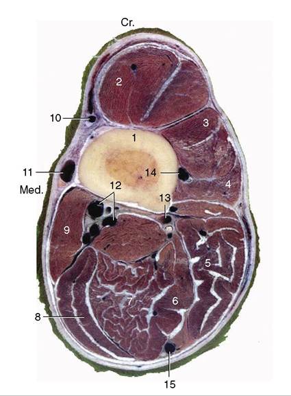
Figure 23-41 Transverse section of the right forearm at the level shown in Figure 23-42.
1, Radius; 2, extensor carpi radialis; 3, common digital extensor; 4, lateral digital extensor; 5, ulnaris lateralis; 6, deep digital flexor; 7, superficial digital flexor; 8, flexor carpi ulnaris; 9, flexor carpi radialis; 10, accessory cephalic vein and medial cutaneous antebrachial nerve (from musculocutaneous); 11, cephalic vein; 12, median artery, veins, and nerve; 13, muscular branches of median vessels; 14, cranial interosseous vessels; 15, ulnar nerve and collateral ulnar vessels.Most veins of the forelimb are satellite, although they are often duplicated or further replicated where they accompany the larger arteries (Figure 23-42/2). Some superficial veins seek independent courses, and those coming from the hoof have already been mentioned. The superficial veins include the cephalic and accessory cephalic veins, which are prominent and palpable in the forearm (Figure 23-42/10,10'). The cephalic vein is joined to the brachial vein via the median cubital at the elbow and continues to ascend in the groove between brachiocephalicus and pectoralis descendens, where it is at risk in “staking” injuries. It joins the external jugular vein at the base of the neck.
Two clusters of lymph nodes drain the free part of the limb. The cubital nodes lie on the medial aspect of the humerus just proximal to the elbow joint. They drain more distal parts of the limb and channel their outflow to the axillary nodes. These lie medial to the shoulder joint in the angle between the axillary and subscapular arteries and drain the arm and shoulder, together with a part of the thoracic wall caudal to the limb. Their efferent vessels go to the caudal deep cervical nodes, and thence the lymph flows directly or indirectly to the veins at the thoracic inlet. The superficial cervical nodes are arranged in a long chain that crosses the deep surface of the omotransversarius and brachiocephalicus (see Figure 18-38/5). The group consists of many small nodes, and because these are embedded in fat and do not form a firm compact mass, the group is not always easily located.
Palpation should be directed to drawing the nodes forward, away from the subclavius against which they lie. The superficial cervical nodes mainly drain skin over the upper part of the limb but also receive some lymph from deeper structures.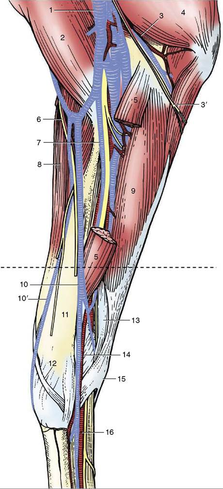
Figure 23—42 Dissection of the medial surface of the right forearm. (The broken transverse line indicates level of section in Figure 23-41.) 1, Multiple brachial veins; 2, biceps; 3, ulnar nerve and collateral ulnar vessels; 3', caudal cutaneous antebrachial nerve; 4, triceps; 5, flexor carpi radialis, resected; 6, medial cutaneous antebrachial nerve; 7, median nerve and vessels; 8, extensor carpi radialis; 9, flexor carpi ulnaris; 10, 10', cephalic and accessory cephalic veins; 11, radius; 12, extensor carpi obliquus; 13, superficial digital flexor; 14, radial artery and vein; 15, accessory carpal bone; 16, medial palmar nerve and vessels.