THE PASSIVE STAY-APPARATUS
It is well-known that horses can remain on their feet for much longer than other domestic animals. In fact, they are thought by many to sleep while standing. This is not quite true: they may rest or doze standing, but for a refreshing sleep they lie down, often only at night when unobserved.
When horses stand quietly, most weight is carried by the tendons, ligaments, and deep fascia of the stay-apparatus, which do not tire; only a minimum of muscular energy is expended.The bony column of the forelimb supports the cranial end of the trunk at the attachment of the serratus ventralis muscle to the medial surface of the scapula (Figure 23-38, A/1). A vertical line dropped from the center of this attachment passes caudal to the shoulder, through the elbow, through or slightly cranial to the carpal joint, and cranial to the fetlock and pastern joints. If unsupported, the column would collapse by flexion of the shoulder and elbow joints, by overextension (or possibly flexion [buckling forward]) of the carpal joint, and by overextension of the fetlock and pastern joints. (The coffin joint actually flexes when the fetlock sinks under weight and can be disregarded in this discussion.)
The shoulder joint is prevented from flexion by the strong internal biceps tendon (Figure 23-38/2) that connects the supraglenoid tubercle of the scapula with the radius. The latter attachment can be regarded as fixed because it is very close to the axis of rotation of the elbow joint (Figure 23-38/5), which is stabilized by the weight on the limb. Tension in the wide biceps tendon puts great pressure on the intertubercular groove of the humerus. Indeed, some believe that the molding of the tendon to the intermediate tubercle actually causes the joint to lock. At its other end, the pull of the biceps is transmitted via the lacertus and extensor carpi radialis (Figure 23-38/6,10) to a second fixed point at the upper end of the large metacarpal bone.
This pull augments the action of the extensors of the carpal joint and prevents that joint from buckling forward and collapsing the limb; any tendency toward overextension is prevented by close packing of the carpal bones in front and by the strong palmar carpal ligament behind (Figure 23-15, A-B/7).The fetlock joint is prevented from overextension principally by the suspensory apparatus (comprising the interosseus, proximal sesamoid bones, and distal sesa- moidean ligaments), which is tensed under load (Figure
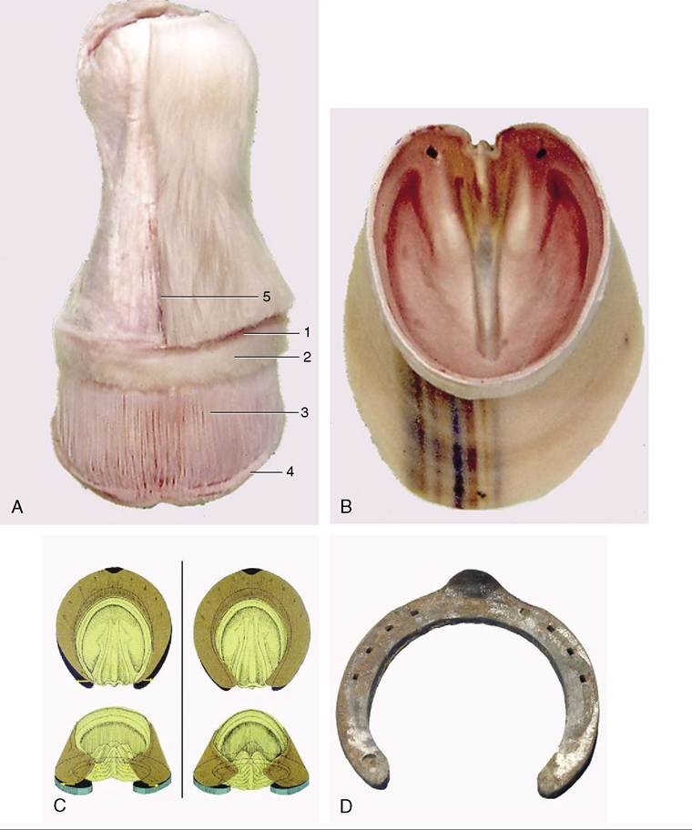
Figure 23-34 A, Dermis exposed by removal of the hoof. B, Hoof shoe removed from specimen A. C, Changes in the form of the hoof during locomotion. D, Shoe showing the heel part polished by movement of the hoof heel. 1, Perioplic dermis; 2, coronary dermis; 3, laminar dermis; 4, terminal papillae on the ends of the dermal laminae; 5, cut edge of skin.
23-38/13,14,16-18). The effect is reinforced by tension in the accessory (check) ligaments and distal parts of the superficial and deep flexor tendons (Figure 23-38/9,11).
Tension in the deep flexor tendon tends to flex the coffin joint, which causes the toe of the hoof to dig into the ground. The extensor branches of the interosseus (Figure 23-38/15), pulling on the extensor process of the bone at impact, counteract this and keep the hoof level.
Overextension of the pastern joint is opposed by the axial and abaxial palmar and straight sesamoidean ligaments (Figure 23-38/18,19), which span its palmar aspect. The taut deep flexor tendon gives additional support. (Buckling forward is prevented by the superficial flexor that attaches on the palmar aspect of the joint.)
With the shoulder joint fixed (by the biceps tendon), the weight of the trunk rests on the upper end of the nearly vertical radius. Therefore, unless the horse sways
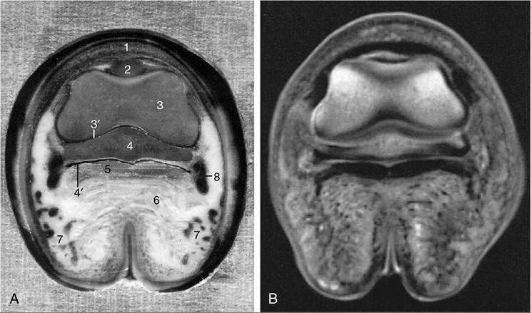
Figure 23-35 A, Transverse section of the digit at the level of the navicular bone, proximal surface.
B, Magnetic resonance image taken at the same level. 1, Coronary dermis; 2, extensor process of distal phalanx (PIII); 3, distal end of middle phalanx (PII); 3', coffin joint; 4, navicular bone; 4, navicular bursa; 5, deep flexor tendon; 6, digital cushion; 7, cartilage of hoof and venous plexus; 8, position of digital vessels and nerve.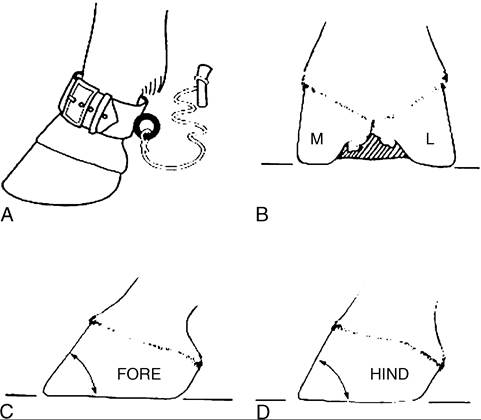
Figure 23-36 A, In former days horses at pasture were hobbled with a "pastern"; this is why the narrow part of the limb above the hoof is known today as the pastern. B, Palmar (plantar) view of the foot; the lateral (L) angle of the wall (with the ground) is more acute than the medial (M). C and D, The angle at the toe is more acute in the forelimb than in the hindlimb.
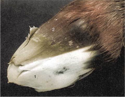
Figure 23-37 Hoof of a newborn foal. 1, Mass of soft, primary horn covering the ground surface and distal half of the hard, permanent hoof wall; 2, pigmented permanent hoof wall.
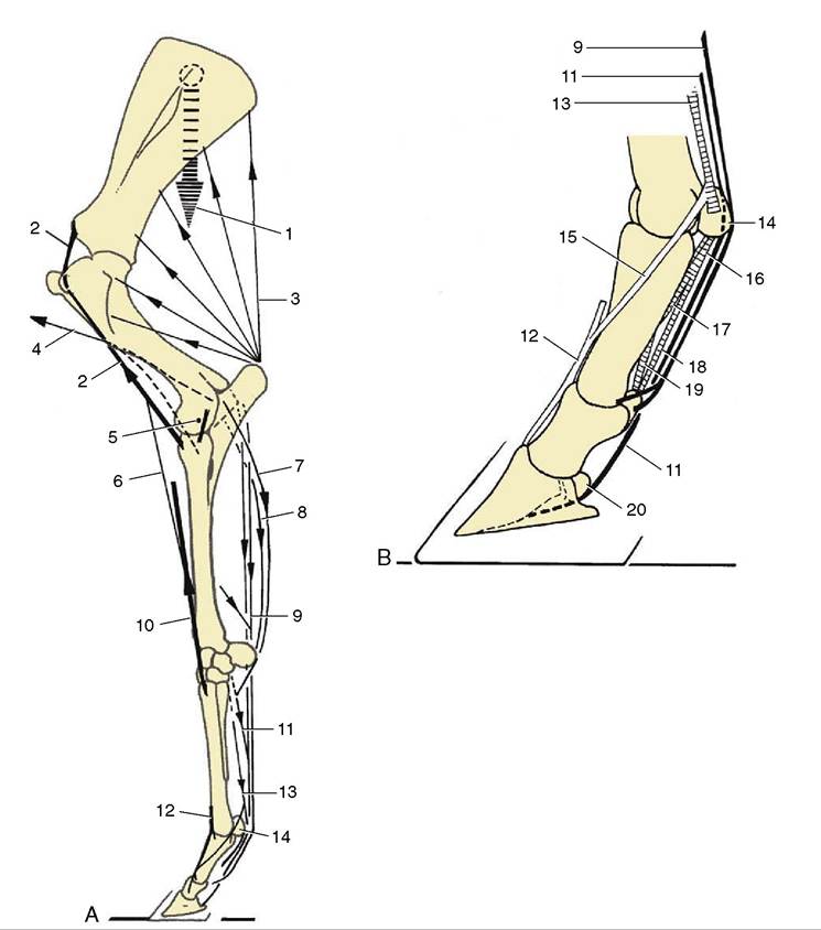
Figure 23-38 A, The stay-apparatus of the left forelimb; lateral view. B, Detail of digit; lateral view. 1, Weight of trunk; 2, internal biceps tendon; 3, triceps; 4, brachiocephalicus and brachial fascia to elbow joint; 5, axis of elbow rotation, next to eccentric collateral ligament; 6, lacertus fibrosus; 7, ulnaris lateralis; 8, flexor carpi ulnaris; 9, superficial digital flexor and accessory (check) ligament; 10, extensor carpi radialis; 11, deep digital flexor and accessory (check) ligament; 12, common digital extensor; 13, interosseus; 14, proximal sesamoid bones; 15, extensor branch of interosseus; 16, 17, 18, cruciate, oblique, and straight sesamoi- dean ligaments; 19, axial palmar ligament; 20, navicular bone.
markedly forward, only small forces are required to prevent the elbow joint from flexing. These are mainly supplied by passive tension of the tendinous components of the carpal and digital flexors (the superficial digital flexor especially) and the eccentrically placed collateral ligaments (Figure 23—38/5,7-9). Recent information indicates that because of their muscle fiber composition—characteristic of postural muscles—the anconeus and the medial head of the triceps may also oppose flexion of the elbow joint. The large mass of the long and lateral heads of the triceps—the principal extensor of the elbow joint—remains flaccid even when the other forelimb is picked up to make the horse stand three-legged (see Figure 23—2/1, and the effects of radial paralysis on page 621).