The Electroencephalogram Is a Common CIinicaITooI
It has been known since the 1930s that a fluctuating electrical voltage reflecting brain activity could be recorded from macroelectrodes on the scalp (Figure 16-2). Such a recording
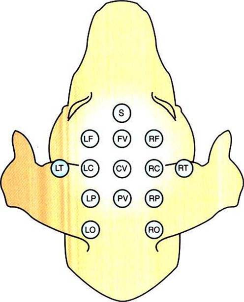
FIGURE 16-2 Commonly used points of scalp electrode attachment (electrode montage) for recording the electroencephalogram (EEG).
Recordings are typically made from multiple paired combinations of electrodes.is known as an electroencephalogram (EEG). The frequency of the waveform recorded varies inversely with its amplitude. Both frequency and amplitude change with changes in levels
of arousal (Figure 16-3). An alert animal has a fairly high- frequency, fairly low-amplitude EEG, whereas a more relaxed animal has a slower-frequency, higher-amplitude EEG. A sleeping animal usually begins sleep by exhibiting a slow-wave, high-amplitude EEG. Paradoxically, there are periods of high- frequency, low-amplitude EEG during the sleep cycle. Four frequency ranges have been given names: alpha (8-13 Hz), beta (13-30 Hz), delta (0.5-4 Hz), and theta (4-7 Hz).
This technique has been applied clinically since the early 1960s. Abnormal EEG activity has been associated empirically with several brain diseases. In human neurology, EEGs have been used to classify the epilepsies, to localize lesions, and to help define “brain death.” EEGs have not been as widely used in veterinary medicine but still have clinical utility in veterinary neurology.
We now discuss where these scalp recordings originate and how they relate to brain function.