THE DENTITION
The dentition of the horse is admirably suited to a diet of grass, a surprisingly abrasive material. The masticatory area is increased by the enlargement of the premolars and their assimilation to the molars with which they present a continuous grinding surface.
Both cheek teeth and incisors have high crowns, which ensure a long working life, despite the considerable attrition that takes place at the occlusal surfaces. Delayed formation of the roots also allows the cheek teeth to grow for some years after they come into wear. Attrition wastes the cheek tooth by 2 to 3 mm each year; to allow for this the greater part of the crown is initially embedded within the jaw and only gradually extruded to compensate for this loss. The enamel casing of the incisor and cheek teeth is also folded, although in different ways in the incisor, upper cheek, and lower cheek teeth series. The folding increases the area of the durable enamel presented at the working surface, where it stands proud of the neighboring dentine; the alternation of harder and softer tissues provides efficient grinding instruments (see Figure 18-19).The formula of the temporary dentition is
and that of the permanent dentition is
The incisor teeth are ranked together to form a continuous arch in each jaw and are so implanted that their roots converge (Figure 18-17). Each is curved lengthwise, presenting a labial convexity. When in occlusion, the upper and lower incisors of the young animal form a continuous arch when viewed in profile. Later, as they wear, the upper and lower teeth meet at an increasingly pronounced angle. The occlusal surface recently brought
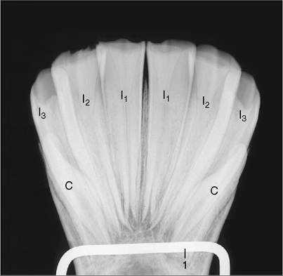
Figure 18-17 Root convergence of permanent lower incisors; radiograph of a bone specimen from a 5-year-old (estimated) horse.
Note the funnel-shaped infundibulum visible in each of the first and second incisors. I1,12, and I3, Lower first, second, and third incisors; C, lower canine tooth, present only in the male; 1, mounting wire of specimen.into use is a broad transverse oval (Figure 18-18, B) and presents an outer enamel casing and an inner enamel ring lining the infolding known as the infundibulum; this is partially filled with cement, leaving a small cavity, the cup (Figure 18-18/7). Because the enamel lining is more resistant, it projects above the surrounding dentine. Changes in the appearance of the occlusal surface provide the information principally used in aging older horses. The points to note are the depth of the infundibulum and its overlap with the dental cavity. Although it may appear that wear would eventually expose the pulp, this is prevented by the timely formation of secondary dentine, distinguishable from primary dentine by its darker color; this secondary dentine provides the feature known as the dental star (Figure 18-18/5).
Although canine teeth generally form in both sexes, they are rudimentary and commonly fail to erupt in mares. In male animals they are low, laterally compressed cones placed within the diastemas rather closer to the corner incisors than to the cheek teeth. The embedded portions are disproportionately large in relation to the exposed crowns.
The first premolar (“wolf” tooth) often fails to develop, and when present, it is vestigial and almost invariably confined to the upper jaw. Although it is without functional significance, it does have a potential nuisance value because it may shift under the pressure of the bit and so irritate the gum. It is easily extracted.
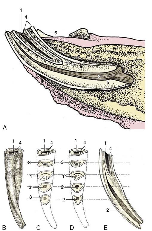
Figure 18-18 Structure of a lower incisor. A, In situ, sectioned longitudinally; the clinical crown is short in relation to the embedded part of the tooth.
B, Caudal view; the junction between the clinical crown and the rest of the tooth is not marked. C, As a result of wear the occlusal surface changes; the cup gets smaller and disappears, leaving, for a time, the enamel spot; the dental star appears and changes from a line to a large round spot. D, These are sawn sections of a young tooth for comparison. E, Longitudinal section of incisor, showing the relationship between the infundibulum and dental cavity; the latter is rostral. 1, Cup, black cavity in center of infundibulum; 1', enamel spot, proximal end of infundibulum; 2, dental cavity; 3, dental star, changing in shape from a linear to a rounded form; 4, outer and inner enamel rings; 5, cement; 6, lingual surface.The remaining premolars (P2-P4) form a continuous row with the molars. The first and last of the six cheek teeth are somewhat triangular in section, the others rectangular; nonetheless, each is so like its neighbors that only an expert may distinguish isolated teeth (see Figure 18-21). There are, however, important differences between the upper and lower sets; the upper teeth are much wider and exhibit a more complicated enamel folding, which creates two infundibula that fill with cement before eruption. The enamel of the lower teeth is also much folded but forms no infundibula (Figure 18-19, B). Most teeth occlude with two members of the opposing set along a relatively narrow area of contact that follows the lingual edge of the upper teeth and buccal edge of the lower teeth. The occlusal plane slopes Ventrobuccally (see Figure 18-10). Irregular or incomplete chewing movements may cause the buccal edge of the upper cheek teeth and the lingual edge of the lower cheek teeth to escape wear (sharp teeth); the resulting
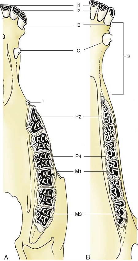
Figure 18-19 The permanent teeth of the upper (A) and lower (B) jaws. 1, "Wolf" tooth (P1); 2, diastema.
protrusions must be filed down (floated) to prevent injury to cheeks and tongue.
The structure of the cheek teeth is shown in Figure 18-20. The upper teeth are anchored by three or four roots and are so implanted that the reserve portions slope caudally at varying angles (Figure 18-21). The relationship to the maxillary sinuses and other features of the skull is very helpfully revealed in radiographs. Only a thin plate of alveolar bone separates the molars from the sinus; in consequence, infection may easily spread to the sinus from tooth or alveolar abscesses. The relationship changes with age, partly because gradual extrusion lowers the alveolar floor, enlarging the sinus, and partly because the teeth migrate rostrally (see Figure 18-15).
The transitory swellings occasionally seen on the ventral margin of the mandible of 2- to 4-year-old horses are produced by modeling of the mandible to accommodate the formation of the roots of permanent teeth, which are prevented from rising within the jaw by remnants (caps) of deciduous predecessors blocking the way (Figure 18-22). When the remnants are shed, their successors can move into place. Further modeling of the mandibular border erases the swellings.
Simple extraction of cheek teeth is more or less impossible. Their length, curvature, and close fit would
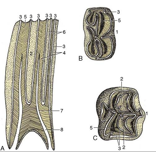
Figure 18-20 Structure of the cheek teeth shown in sagittal section (A) and by views of the occlusal surface of lower (B) and upper (C) molars. 1, Buccal (labial) surface; 2, infundibulum; 3, enamel; 4, dentine; 5, secondary dentine; 6, cement; 7, dental cavity; 8, root canal.
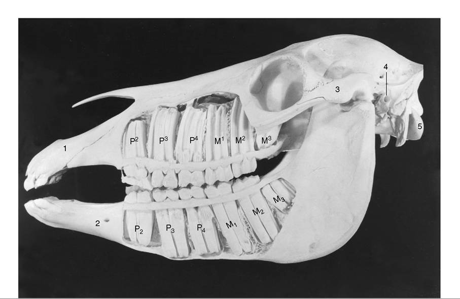
Figure 18-21 Exposed cheek teeth of a horse 21/2 years old (estimated). Upperjaw:The deciduous premolars are still present, p2 in the form of a cap; M3 has not yet erupted. Lower jaw: The deciduous premolars 3 and 4 are still present in the form of caps; M3 has not yet erupted.
1, Incisive bone; 2, mental foramen; 3, zygomatic arch; 4, external acoustic meatus; 5, occipital condyle.hamper any effort to draw one out past its neighbor(s), even were the attempt permitted by the small size of the opening between the lips and the depth of the oral cavity (Figure 18-23). Instead, they must be removed by expulsion, that is, by means of a punch brought to bear over the root in an operation of some severity and difficulty involving the opening of a window through bone. Accurate determination of the position of the root of the tooth involved is essential, and for this it is necessary to be mindful of how the dispositions of the teeth change with age. The approach to a caudal member of the upper cheek teeth series is made via the caudal maxillary sinus or the frontal and caudal maxillary sinuses when M3 is involved.
The deciduous teeth generally resemble the permanent teeth but are much smaller and significantly shorter in relation to their breadth. The deciduous incisors are constricted at the neck and are much whiter than their replacements because the porcelain-like enamel is unobscured by the cement encrustation that gives permanent teeth a slightly yellow and porous appearance. Some longitudinal striation is apparent on the temporary incisor crown.