THE EYE AND ORBIT
The margins of the orbit are easily palpable. They are formed by the frontal, lacrimal, and zygomatic bones, with the gap in the dorsolateral segment closed by the orbital ligament (Figure 11-11/d).
Only the medial third of the orbital wall is osseous; the remainder is provided by the periorbita. The orbital axis takes a dorsal, lateral, and anterior direction from the apex of the cone. In brachycephalic dogs, particularly those with wide skulls, the axes point more laterally, restricting binocular vision.The openings into the orbit comprise the optic canal, orbital fissure, duplicated ethmoidal foramina, and the fossa of the lacrimal sac. The optic canal transmits the optic nerve and internal ophthalmic artery; the orbital fissure transmits the oculomotor, trochlear, abducent, and ophthalmic nerves; the ethmoidal foramina transmit divisions of the like-named nerve and artery; and the fossa contains the slight enlargement at the origin of the nasolacrimal duct.
The osseous wall of the orbit is related dorsomedi- ally to the frontal sinus and rostromedially to the maxillary recess; infection in either of these cavities can easily spread to orbital structures. The periorbita is related as follows: medioventrally to the medial pterygoid muscle; ventrally to a pad of fat caudal to the orbital margin, the zygomatic gland, and the large deep facial vein; laterally to the zygomatic arch; and caudodorsally to the orbital ligament and temporalis muscle. The dorsolateral aspect of the orbit is accessible to surgery without resection of bone.
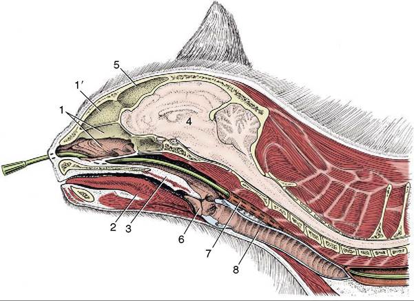
Figure 11-35 Paramedian section of the feline head and neck. A nasogastric tube is in place. 1, Nasal cavity; 1', dorsal part of nasal cavity; 2, tongue; 3, soft palate; 4, brain; 5, frontal sinus; 6, epiglottis; 7, esophagus; 8, trachea.
The important maxillary artery and nerve and their branches to the face and palate course ventral to the orbit between the medial pterygoid and the zygomatic gland (Figure 11-36). The maxillary artery gives off the external ophthalmic artery, which pierces the periorbita near its apex to supply structures within the cone. The temporalis, which surrounds the coronoid process of the mandible, impinges on the periorbita when the mouth is opened. This may cause pain when the orbital contents are diseased as, for example, by a retrobulbar abscess. The proximity to the oral cavity permits drainage of such abscesses into the mouth, behind the last cheek tooth.
The dimensions of the orbital rim of large and small dogs differ less than might be expected; because the diameter of the eyeball varies even less, the surgical working “space” is generally narrower in larger dogs. However, the position of the eyeball within the orbit differs markedly. In dolichocephalic dogs the eyeball is deeply placed and the palpebral fissure is small. The eyes of brachycephalic dogs protrude and are more susceptible to injury to the cornea.
The lacrimal gland (Figure 11-36/7) is flat, lobulated, and about 12 to 15 mm in width. It lies between the eyeball and the orbital ligament, dorsal to the lateral angle of the eye. The gland must be identified and removed in enucleation (removal) of the eye. The thin edge of the third eyelid is visible in the medial angle of the eye in the “resting” state. More is seen when the upper and lower lids are retracted with the fingers, while full protrusion is obtained by gentle pressure on the eyeball through the upper lid (see Figure 9-21/6). Although the superficial gland that surrounds the cartilage of the third lid is not normally visible, it appears when the eyelid is retracted because the increased retrobulbar pressure pushes it to the fore. Active protrusion of the third eyelid, effected by a specific muscular arrangement, is common in cats and may have an emotional or physical origin.
Abnormal retrobulbar pressure may cause the gland of the third eyelid to be everted into the medial angle of the eye, where it appears as a round swelling below a covering of conjunctiva. Sub- epithelial lymph nodules present on the bulbar surface of the third eyelid may become inflamed.In cross section the eyelids display the external skin, the orbicular muscle of the eye, the tarsal plate, the meibomian glands, and the palpebral conjunctiva. The openings of the tarsal glands (20 to 40 in each lid) can be seen at the lid margins. When the lids are everted, these glands appear as white cords extending 5 to 7 mm from the lid margin under the conjunctiva. Occasionally aberrant hairs protrude from the openings of the tarsal glands and may irritate the cornea. The eyelashes in dogs are found on the outer surface of the upper lid margin; there are none on the lower lid. Both lids of cats are without lashes.
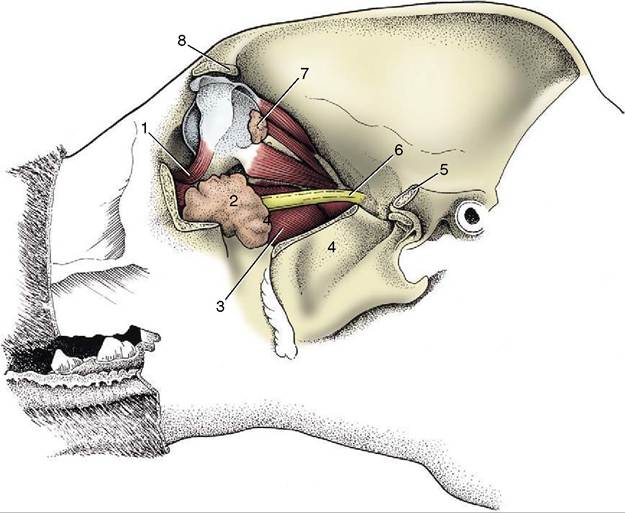
Figure 11-36 Dissection of the canine orbit and pterygopalatine fossa, lateral view. 1, Ventral oblique muscle; 2, zygomatic gland; 3, medial pterygoid muscle; 4, coronoid process of mandible, cut; 5, caudal stump of zygomatic arch; 6, maxillary nerve; 7, lacrimal gland; 8, zygomatic process of frontal bone.
The orbicular muscle of the eye, rostral to the tarsal plate, is anchored to the orbit by fascia medially and by the retractor muscle of the lateral angle laterally. These attachments preserve the elliptical shape of the palpebral fissure.
The puncta lacrimalia are 2 to 4 mm from the medial angle of the eye and are usually located at the junction of pigmented and nonpigmented epithelia. Although they may be difficult to find or the lower one may in fact be absent or displaced to the bulbar surface of the lid, it is possible to cannulate them. The puncta are the openings to the upper and lower canaliculi, which join to form the lacrimal sac, from which the nasolacrimal duct takes origin (see Figure 9-21).
The duct continues rostrally in the medial wall of the maxilla, deep to the nasal mucosa. An accessory, or more rarely the sole, opening of the nasolacrimal duct may enter the nose at the level of the canine tooth in a significant proportion of dogs. The duct makes an abrupt 90° turn about 2 mm before opening onto the floor of the nasal cavity (see Figure 11-8).The feline lacrimal system is similar; however, an opening with the oral cavity has been recorded, located on a small papilla just behind the upper incisor teeth.
One or both puncta may be absent in several dog breeds, as well as Persian cats. If both are absent, a slight depression in the conjunctiva may indicate where the opening would normally have been located.
The eyeball is nearly spherical and relatively large. The cornea is slightly oval, its larger diameter being mediolateral in keeping with the shape of the globe itself. It is slightly thicker at the pole than at the periphery. The canine iris is brown, golden yellow, or bluish, and whether dilated or contracted the pupil remains round. It is said to be smaller in older dogs under standard light conditions. Remnants of the papillary membrane may be seen on its upper margin in puppies up to the age of 5 weeks.
The fundus is illustrated in Figure 11-37, A-B. The triangular tapetum lucidum, which nearly fills the dorsal half, includes the optic disc in large dogs. The retinal vessels radiate from the disc; prominent venules form a partial circle from which tributaries usually spread dor- sally, medioventrally, and lateroventrally. Thinner arterioles extend in all directions, many accompanying the venules.
In the cat there is little surgical working space between the eye and the orbital margin. The third eyelid is large, and in certain circumstances it may be drawn completely over the cornea. As in the dog, it responds to retraction of the eyeball. The cornea is relatively large and permits a wide visual field. The color of the iris ranges from blue through green to golden.
In certain breeds iris color is strictly prescribed to meet show standards. Kittens are usually born with blue eyes that later change color.The pupils of domestic cats are round when dilated but are vertical slits when constricted (those of some wild felids remain round at all times) (Figure 11-38, A-B). The vertical form is due to the dorsoventral orientation of muscle fibers that extend to the periphery
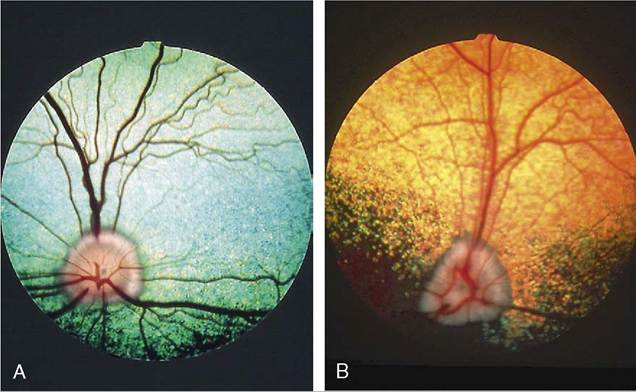
Figure 11-37 Fundus of eye. A, Dutch Sheepdog. B, Old English Sheepdog.
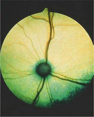
Figure 11-39 Fundus of eye in a cat.
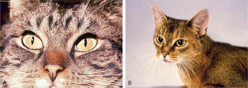
Figure 11-38 A, Slit form of constricted feline pupil. B, Round form of dilated feline pupil.
of the iris and decussate at the extremities of the pupil. The fundus is dominated by a large tapetum lucidum that surrounds the optic disc. The tapetum is yellowish- or bluish-green and because of its brilliance is thought to be more effective in reflecting light than that of the dog, which may be a convenience in nocturnal wandering (Figure 11-39).