The Fetal Circulation Mixes Oxygenated and Deoxygenated Blood at Several Points, So the Fetus Exists in a State of Hypoxemia
In the adult the cardiac output of the right and left ventricles is separate and perfuses the pulmonary and systemic circulations, respectively. In the fetus the output of the two sides of the heart mixes at several points, so it is convenient to use the term cardiac output to refer to the combined output of the right and left ventricles.
The combined cardiac output averages 500 mL/min/kg in fetal sheep; the output of the right ventricle exceeds that of the left (Figure 51-4).The placenta, which has a low vascular resistance, receives 45% of the cardiac output through the umbilical arteries. The umbilical veins drain the placenta toward the liver. In species such as the sheep, most of the umbilical venous blood passes through the liver through a low-resistance channel known as the ductus venosus; in other species, such as the pig and horse, the ductus venosus disappears early in gestation, and umbilical venous blood flows through the liver capillaries. Within the liver, the oxygenated blood from the placenta is mixed with a small amount of more poorly oxygenated blood draining the liver sinusoids. The hepatic venous blood enters the posterior vena cava, where it mixes with poorly oxygenated blood, draining the hind end of the fetus, so the blood returning to the right atrium has a Po2 of 25 mm Hg.
A low-resistance pathway, the foramen ovale, connects the right and left atria, and a structure known as the crista dividens directs the better-oxygenated blood from the posterior vena cava through the foramen ovale to the left atrium. The poorly oxygenated blood returning to the right atrium in the cranial vena cava is directed into the right atrium and right ventricle. Most of the output of the right ventricle does not go through the lungs, however, because fetal lungs have a high vascular resistance. Another low-resistance channel, the ductus arteriosus, connects the pulmonary artery with the aorta and allows blood to bypass the lungs. It is important to note that the arrangement of the fetal circulation allows the better-oxygenated blood to enter the left ventricle, from which it reaches the brachycephalic vessels and the front of the animal.
The more poorly oxygenated blood from the ductus arteriosus enters the aorta downstream from the brachycephalic vessels. The tissues of the hind end of the animal and the placenta receive blood with a Po2 of approximately 22 mm Hg.Flow of blood from the right atrium to the left atrium through the foramen ovale and from the pulmonary artery to
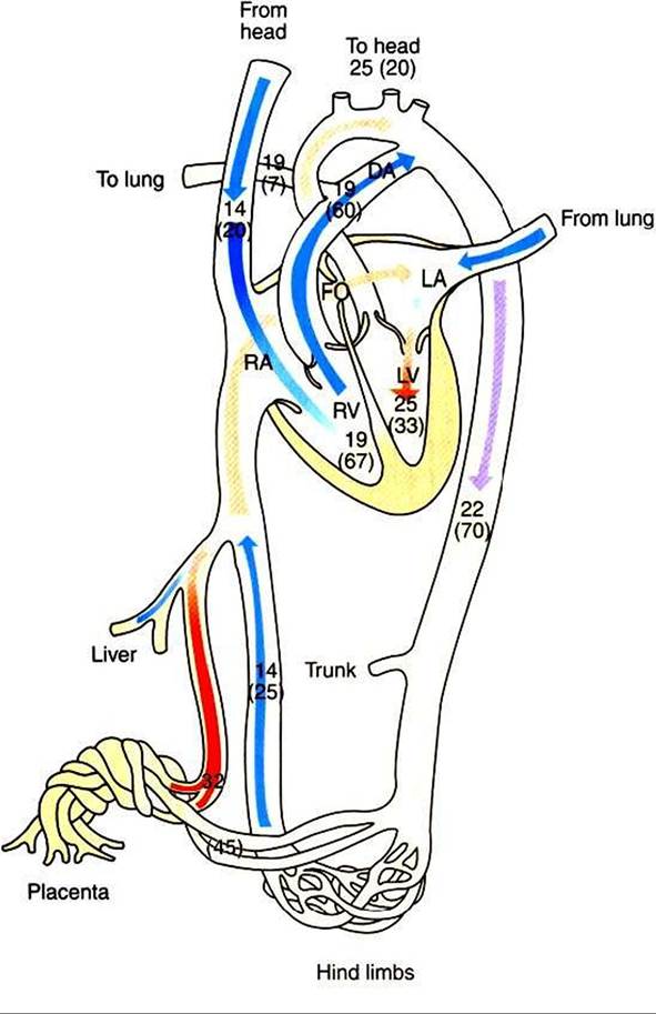
FIGURE 51-4 Diagrammatic representation of the fetal circulation showing oxygen tension (Po2) in millimeters of mercury (mm Hg) and percentage of cardiac output (in parentheses) in different parts of the circulation. DAt Ductus arteriosus; FO, foramen ovale; LA, left atrium; LV, left ventricle; RA, right atrium; RV, right ventricle.
the aorta through the ductus arteriosus requires that the pressure in the right side of the fetal circulation be greater than that in the left side. This pressure difference occurs because the left side of the circulation provides most of its output to the low-resistance placenta, whereas the right side of the fetal circulation is opposed by the high-resistance pulmonary circulation. At term, systemic arterial pressure in the lamb is about 42 mm Hg.
The fetal circulation is not a passive system and is capable of considerable regulation, particularly as the fetus matures. Fetal hypoxia can stimulate vasodilation in the heart and brain and vasoconstriction in the gut, kidneys, and skeletal tissues. The fetal pulmonary circulation constricts vigorously when the fetus is hypoxic. This constriction diverts more blood through the ductus arteriosus to the systemic tissues.