THE GUSTATORY ORGAN
The receptors for the sense of taste are the taste buds (Figure 9-32), microscopic nests of cells mainly associated with the papillae of the tongue, although small numbers are also found in the soft palate and in the vicinity of the epiglottis.
Taste buds are about as tall as the epithelium in which they lie and communicate with the oral cavity by taste pores through which solutions enter to stimulate the receptor cells. Taste pores cannot be seen with the naked eye.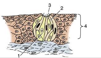
Figure 9-32 Histological section of a taste bud. 1, Sustentacular cell; 2, gustatory cells; 3, taste pore; 4, epithelium.
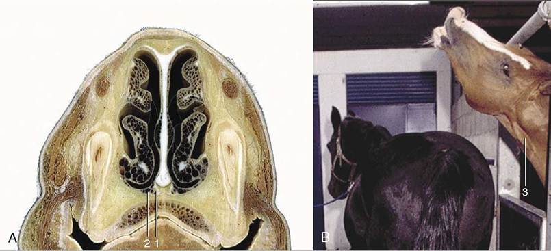
Figure 9-31 A, Transverse section of vomeronasal organ of horse. B, During the Flehmen reaction the head is fully extended, accentuating several features of the neck. 1, Vomeronasal cartilage; 2, vomeronasal duct; 3, jugular groove.
The taste buds consist of sustentacular or supporting cells in addition to the receptor or gustatory cells. The latter have elongated nuclei and at their free tips bear microvilli (taste hairs) that project into the taste pore. Glands deep to the papillae discharge a serous secretion on the surface of the epithelium. It is believed that the secretion cleanses the taste pores and enhances perception by the gustatory cells.
To be discerned, food substances have to be in solution. One of the reasons food is insalivated is to dissolve parts for sampling by the taste buds. The principal taste sensations are sweetness, sourness, and saltiness. In the dog it appears that sweetness and saltiness are perceived in the rostral two thirds of the tongue where taste buds are present on the fungiform papillae. Sour substances are perceived over the entire tongue. The caudal third of the tongue, which incorporates the vallate and foliate papillae, therefore seems to respond only to what tastes sour.
The afferent pathways mediating these sensations are similarly divided. In the rostral two thirds of the tongue the pathways travel at first in the lingual nerve and then in the chorda tympani, which we encountered in the description of the ear. After passing through the geniculate ganglion of the seventh cranial nerve, they enter the medulla oblongata. The afferent fibers from the caudal third of the tongue travel in the glossopharyngeal nerve (and to a small extent in the vagus) to the medulla oblongata.