THE KIDNEYS
The kidneys have the maintenance of the milieu interi- eur as their prime task. They do this by filtering the
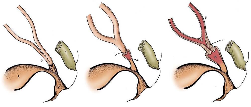
Figure 5-15 The fusion of the combined paramesonephric ducts with a bud from the urogenital sinus forms the vagina.
1, Rectum; 2, caudal part of urogenital sinus (vestibule); 3, cranial part of urogenital sinus (bladder, urethra); 4, bud from urogenital sinus; 5, fused paramesonephric ducts; 6, vagina; 7, cervix uteri; 8, uterine horn.
Figure 5-16 Different degrees of fusion of the paramesonephric ducts. A, Uterus duplex (rabbit). B, Uterus bicornis (small body: sow, cow). C, Uterus bicornis (large body: mare). D, Uterus simplex (woman). 1, Infundibulum; 2, uterine tube; 3, uterine horn; 4, fusion site of the two ducts; 5, cervix; 6,
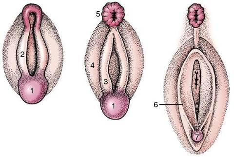
Figure 5-17 Development of the female external genitalia. 1, Genital tubercle; 2, cloacal fold; 3, urogenital fold; 4, lateral swelling; 5, anus; 6, labia of vulva; 7, clitoris.
vagina; 7, vestibule.
plasma, initially extracting an enormous volume of fluid before subjecting this ultrafiltrate to further processing in which useful substances are selectively reabsorbed, waste substances are concentrated for elimination, and the volume is adjusted by the conservation of sufficient water to maintain the composition of the plasma within the appropriate range. Some figures may give an impression of the dimensions of this task. In large dogs (and animals of similar size), 1000 to 2000 L of blood perfuse the kidneys daily; the 200 to 300 L of fluid that are filtered from this volume are later reduced by reabsorption until only 1 or 2 L of urine remain to be discharged.
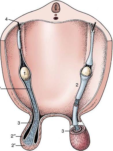
Figure 5-18 Schematic representation of the testis and gubernaculum within the peritoneal fold in which descent takes place.
1, Testis; 2, gubernaculum; 2', pars propria; 2'', pars infravaginalis; 2'", pars vaginalis; 3, vaginal process; 4, testicular artery.The endocrine function of the kidneys consists of the production and release of two hormones: renin, which plays a vital role in the regulation of systemic blood pressure, and erythropoietin, which influences erythropoiesis. Both are produced within the juxtoglomerular complexes, localized regions of intimate association between arterioles formed by the union of afferent glomerular capillaries with adjacent portions of the distal convoluted tubules (p. 222).
The kidneys are firm, reddish-brown glands whose appearance varies considerably among mammals (Figure 5-21). The most familiar form, that which has introduced the term kidney-shaped to the common vocabulary, is encountered in the dog (Figure 5-21, D), cat, and small ruminants. The kidneys of the pig (Figure 5-21, C) are a much flattened version, whereas those of the horse (Figure 5-21, E) are more heart-shaped. In contrast, the bovine kidneys (Figure 5-21, B) are very dissimilar and have a surface deeply fissured to outline many lobes. Even greater subdivision is shown by the kidneys of certain marine species (Figure 5-21, A), which resemble trusses of grapes that have the lobes only slightly fused and mainly held together by the branching “stalk.”
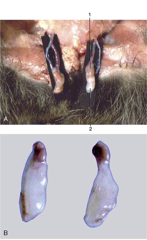
Figure 5-19 Stages in the process of gubernacular swellings. The testis and gubernaculum have already passed the inguinal canal. Inguinal area of newborn pup. A, 1, Testis; 2, exposed gubernaculum. B, Testis and gubernaculum of pig fetus (110 days).
The kidneys are usually found pressed against the abdominal roof, one to each side of the vertebral column, and predominantly in the lumbar region, although often extending forward under the last ribs. Their positions change with the excursions of the diaphragm, and they move, perhaps by half the length of a vertebra, with each breath.
They are rarely symmetrical; in domestic animals, other than pigs, the right one is about half a kidney-length in advance of its fellow. The cranial extremity of the right kidney commonly fits into a fossa of the liver, which helps fix its position. The left one, lacking this lodgment, is more mobile and is more likely to sag within the abdomen. The pendulous left kidney of ruminants is thrust into the right half of the abdomen by the enormous development of the stomach. In general, kidneys pressed against the abdom-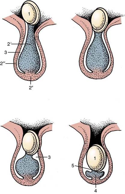
Figure 5-20 Successive stages in gubernacular regression in the pig fetus. Observe the migration of the testis caused by this regression. 1, Testis and epididymis; 2, gubernaculum; 2', pars propria; 2", pars infravaginalis; 2'", pars vaginalis; 3, vaginal process; 4, ligament of the tail of the epididymis; 5, proper ligament of the testis.
inal roof are largely retroperitoneal, whereas those suspended at a lower level have a more extensive peritoneal covering (Figure 5-22).
Each kidney lies within a splitting of the sublumbar fascia, which also holds considerable fat (sometimes enough to hide the kidney completely). The fat protects against distorting pressures from neighboring organs. The surface of a kidney is generally smoothly convex except for an indentation of the medial border. This indentation leads to a concealed space (renal sinus; Figure 5-23) occupied by the dilated origin (renal pelvis) of the ureter, the vessels and nerves passing to and from the renal hilus, and more fat.
The general organization of the kidney is most conveniently shown in a section that divides the organ into dorsal and ventral “halves.” Such a section shows that the parenchyma is enclosed within a tough fibrous capsule. The capsule restricts the kidney’s ability to expand; the swelling that occurs in certain disease conditions therefore tends to compress the tissue and narrow the internal passages.
The capsule strips readily from the healthy kidney but adheres where the underlying substance has been scarred by former lesions.The parenchyma is visibly divided into an outer cortex and an inner medulla (see Figure 5-23). The cortex is distinguished by its reddish-brown color and finely granular appearance. The medulla consists of a dark, purplish outer zone, from which stripes (medullary rays) extend into the cortex, and a paler, grayish- red, and radially striated inner zone that extends toward the renal sinus. The gross arrangement of the medulla shows very marked species differences. In many species the medulla is arranged as several (or even many) discrete masses, each roughly pyramidal in form. In kidneys of this type a portion of the cortex is associated with each pyramid and caps its base, the aspect directed toward the outer surface. The apex of the pyramid points toward the renal sinus and forms a papilla that fits into a cuplike expansion (calix) of the renal pelvis. Each medullary pyramid with its associated cortex constitutes a renal lobe. Kidneys that retain this organization are said to be multipyramidal or multilobar. In some multipyramidal kidneys, such as those of cattle (Figure 5-23, A), the boundaries between the lobes are revealed by the fissures that penetrate from the surface; in others, including those of pigs, no external evidence of lobation is present (Figure 5-23, B).
All mammalian kidneys pass through a multipyrami- dal phase in their development, although in most species the number of lobes is later drastically reduced (Figure 5-24). In some species, including the dog, horse, and sheep, all the pyramids finally fuse to form a single medullary mass that confines the cortex to the periphery, where it forms a continuous shell. Even this unipyramidal or unilobar type of kidney retains some evidence of its complex ontogeny; a slight scalloping of the corticomedullary junction, punctuated by the arteries that mark the interlobar boundaries, shows where the pyramids fused.
The fusion joins the papillae in a common crest (Figures 5-25 and 5-26) that may be modeled to reveal its composite origin; it is so modeled in the dog and goat but not in the horse.The functional units within the kidney are known as renal tubules or nephrons. These epithelial tubules are supported by a connective tissue interstitium and are estimated to number several hundred thousand or even a million in canine kidneys. The structure and the functions of the nephron are more appropriately described in texts of microscopic anatomy and physiology; only a few points, mainly those discernible to the naked eye, are mentioned here.
Each nephron begins with a blind expansion that is invaginated by a cluster of capillaries known as a glomerulus (Figures 5-27/1 and 5-28). The glomerulus and
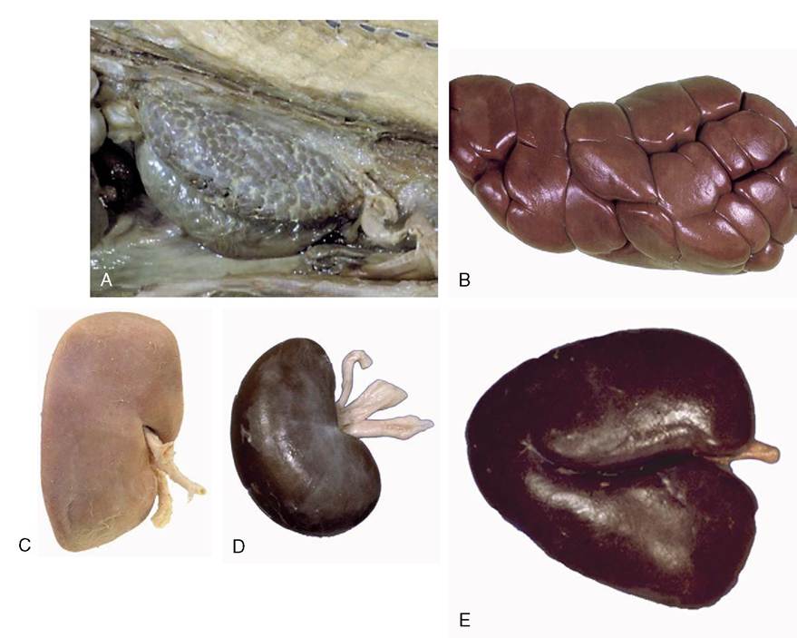
Figure 5-21 Kidney of a dolphin (A), kidney of a cow (B), kidney of a pig (C), kidney of a dog (D), and kidney of a horse
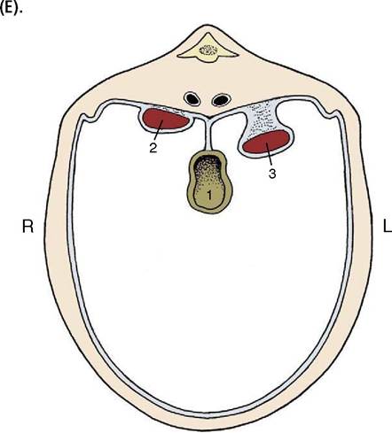
Figure 5-22 Schematic representation of the position of the kidneys in relation to the peritoneal cavity. 1, Gut; 2, right kidney (retroperitoneal); 3, left kidney (intraperitoneal: pendulous or "floating").
its epithelial covering together constitute a renal corpuscle (Figure 5-27Z1'), a structure just large enough to be visible to the unaided eye, especially if the capillaries are congested. The corpuscles are scattered throughout the cortex and give it a finely granular appearance.
The remaining part of the nephron forms a long tubule differentiated into several successive segments. The first, the proximal convoluted tubule, is very tortuous and is located close to the corpuscle from which it arises (Figure 5-27Z2). This part gradually straightens and enters one of the narrow rays that penetrate the cortex from the medulla. The tubule then forms a long hairpin loop (formerly known as the loop of Henle) within the medulla.
The first part of the loop, the descending limb, is relatively narrow and runs through the medulla to approach the papilla before turning back. The ascending limb is generally thicker, although the change in caliber need not coincide with the change in direction, and runs back to regain the medullary ray. On leaving this, the tubule forms a second or distal convoluted part that is also placed close to the corpuscle of origin (Figure 5-27Z4). A short junctional section then runs to join a collecting tubule within the medul-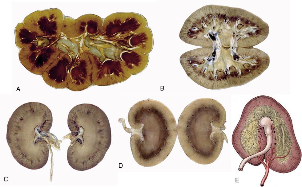
Figure 5-23 Sectioned kidney. Notice that the complexity of the renal pelvis decreases from cow to horse. Cow (plastinated specimen) (A), pig (B), dog (C), cat (D), horse (E).
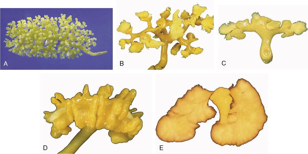
Figure 5-24 Corrosion casts of the renal pelvis of: A, Dolphin, note the branched renal pelvis with many calices. B, Cow, note the papillary ducts extending from the calices. C, Pig, the renal pelvis becomes confluent; again note the papillary ducts. D, Dog, the renal pelvis is one cavity but note the ridges between the renal papilla. E, Horse, one simple renal pelvis and many papillary ducts opening into the renal pelvis.
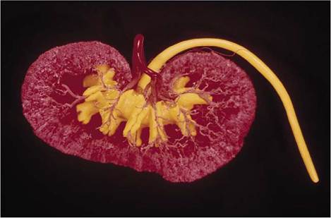
Figure 5-25 Corrosion cast of canine kidney. The renal pelvis and ureter are filled with yellow plastic. Notice the indentations in the pelvis corresponding with the crests of the renal papillae. The ramifications of the renal artery (red) are clearly visible.
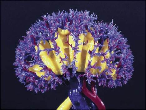
Figure 5-26 Corrosion cast of renal pelvis, renal artery, and renal veins of a goat. The depressions of the ridges of the renal papillae are clearly visible.
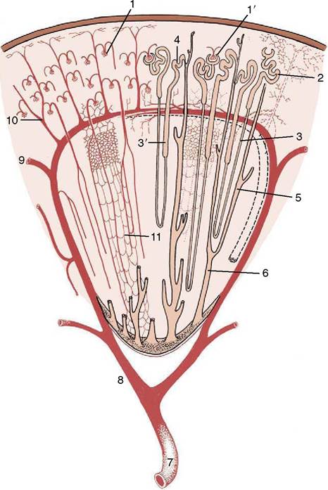
Figure 5-27 Schematic drawing of a kidney lobe. 1, Glomerulus; 1, renal corpuscle; 2, proximal convoluted tubule; 3, descending limb of nephron; 3, ascending limb; 4, distal convoluted tubule; 5, collecting tubule; 6, papillary duct; 7, renal artery; 8, interlobar artery; 9, arcuate artery; 10, interlobular artery; 11, capillary plexus.
lary ray. Each collecting tubule (Figure 5-27/5), which serves many nephrons, runs through the medulla before opening into a larger vessel, a papillary duct, close to the apex (Figure 5-27/6). Several score of papillary ducts drain into the renal pelvis. The papillary ducts can be clearly demonstrated in resin-injection specimens (see Figure 5-24). The perforated (cribriform) areas where they discharge are confined to the apices of independent papillae or to specific regions of a common crest.
Variations in the location of the corpuscles and in the overall length and proportions of the tubules have functional importance that cannot be discussed here.
Each kidney is supplied by a renal artery, a branch of the abdominal aorta, which may carry more than a
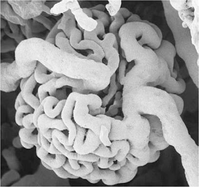
Figure 5-28 Scanning electron micrograph of a corrosion cast of a rat renal glomerulus.
tenth of the total output of the left ventricle! The renal artery divides into several interlobar arteries (Figure 5-27/8) that follow the divisions, former or extant, between the renal pyramids at the corticomedullary junction. These vessels are prominent in gross sections of the kidney. They give rise to branches known as arcuate arteries that curve over the bases of the pyramids (Figure 5-27/9). These in turn give origin to numerous interlobular arteries that supply the units or lobules into which the cortex is divided by the medullary rays (Figure 5-27/10). Each interlobular artery gives rise to many branches that supply individual glomeruli. The glomerular capillaries rejoin in one emissary vessel at the distal pole of the glomerulus, and this then supplies a further capillary plexus around the tubules (Figure 5-27/11). The flow of blood through this second capillary bed is countercurrent to the direction of the urine flow. The vessels that issue from the juxtomedul- lary corpuscles (those in the innermost layer of the cortex) have a particular importance in the supply of the medulla. The renal circulation is actually more complicated than is described here and provides opportunities for collateral circulation. However, the interlobular arteries are certainly functional end-arteries, and the interlobar arteries are possibly functional end-arteries.
The veins, which lead ultimately to the caudal vena cava, are broadly satellite. Lymphatic vessels drain to nodes of the lumbar series that accompanies the aorta. The sympathetic nerves to the kidneys are routed through the celiacomesenteric plexus and thence along the renal arteries. The synapses may be located within the major ganglia or within smaller (aorticorenal) ganglia within peripheral parts of the plexus. The vagus contributes the parasympathetic supply.