» The Lungs (see also pp. 149-156)
The lungs of the dog obtain their distinctive appearance from the deep fissures that divide the lobes, sometimes so completely that they remain connected by little more than the branches of the bronchial tree and pulmonary vessels.
In consequence, torsion of a lobe is a possible complication of thoracic trauma, perhaps most frequently seen after traffic accidents. In contrast, lobulation is not evident to the naked eye through the covering pleura. The right lung, always somewhat the larger, possesses cranial, middle, caudal, and accessory lobes (Fig. 13.11); the left one has only a divided cranial lobe and a caudal lobe. The cardiac impression on the medial surface of the left lung is shallower than that on the right. Despite the existence of a small notch between the two parts of the cranial lobe, the left lung practically covers the lateral face of the pericardium. The notch between the cranial and middle lobes of the right lung is larger and restricted to the ventral part of the fourth intercostal space. It is recommended for heart (right ventricular) puncture and for ultrasonic cardiac imaging.Pulmonary ligaments connect the hilar region of the left lung to the aorta and that of the right lung to the esophagus, which it follows to the hiatus in the diaphragm.
Lung Auscultation
The fields for auscultation and percussion of the lungs are triangular: the cranial border is provided by the 5th rib (actually the caudal border of the triceps), the dorsal border is provided by the lateral margin of the back muscles from the 5th rib to the 11th space, and the basal border is provided by the line joining the 6th costochondral junction, the middle of the 8th rib, and the dorsal end of the 11th space. The forelimb may be drawn forward to increase the accessible area by the space of a couple of ribs.
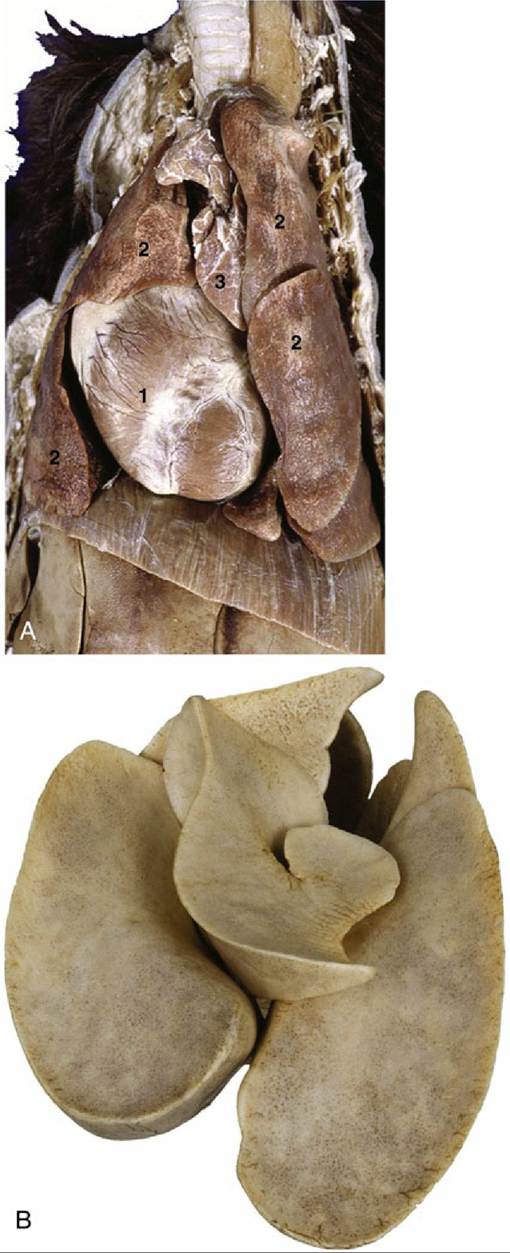
FIG.
13.11 (A) Thoracic viscera of the dog. 1, Heart; 2, pulmonary lobes; 3, thymus. In (B), an inflated specimen, the deep fissures between the lobes of the lung are clearly visible.In plain radiographs, the principal features of the lungs are made by the vessels and bronchi. The blood within the arteries and veins, which cannot be immediately differentiated, produces a pattern of light streaks radiating from the hilar region toward the periphery, branching and tapering as they go. The bronchi, being filled with air, provide dark streaks that contrast less definitely with the lung parenchyma. Bronchial walls may be invisible or may appear as narrow, whitish lines, especially in older animals, in which the cartilage tends to have calcified. The relationships within the bronchial-vascular triads vary in different regions and in different radiographic views. The components are most clearly depicted when portrayed end on; the dark circles of the bronchial lumina are then flanked by white circles representing the companion vessels. The subpleural connective tissue bordering the interlobar fissures may appear as fine lines when penetrated tangentially on a radiograph view.
Both the bronchial tree and the pulmonary vasculature may be made more evident by the use of an appropriate contrast technique (contrast bronchography: Fig. 13.10; angiocardiography: see Fig. 13.23). The larger divisions of the bronchial tree are then very clearly depicted, and if the normal pattern of branching is known, any deviation may reveal the existence of pathology. A more exact picture of the nature and extent of that pathology may be obtained by the use of bronchoscopy, which also requires familiarity with the branching pattern. The principal bronchi produced at the bifurcation of the trachea are separated by a sharp ridge, the carina. The bronchi that initially branch from the principal bronchi supply the different lobes and are named accordingly. The divisions of the next order, the segmental bronchi, also arise according to a consistent pattern and are each associated with well-defined parts of the lobes.
Subsequent divisions into smaller bronchi are less regular and less predictable. The parts of lung associated with the segmental bronchi (the bronchopulmonary segments) constitute the divisions of the lungs on which surgery is based. Various systems of nomenclature such as those based on topography have been devised for the identification.The branching progressively produces a greater cross-sectional area and reduces resistance to the air as it flows into deeper parts of the lung. This process is similar to that in the upper respiratory system, in which the nostrils, the nasal cavity, the pharynx, the larynx, and the trachea each offers successively less obstruction than the preceding segment. It is estimated that the resistance to the inspiratory airflow in dogs is 79% due to the nasal, 6% to the laryngeal, and 15% to the bronchopulmonary parts of the tract; the corresponding figures at expiration are given as 74%, 3%, and 23%, respectively. The resistance to inspiratory and expiratory air in the nasal cavity is further increased in the brachycephalic breeds of dogs, resulting in breathing difficulty even under resting conditions. Except for their shallowness, the lungs of the cat are not significantly different from those of the dog.