THE MAMMARY GLANDS
Examination of one of the several units formed along the trunk of a lactating sow (see Figure 10-31, B) reveals that it is composed of glandular tissue supported and enclosed by a fibrous tissue framework in which run the mammary vessels and nerves.
The whole formation is pervaded with fat and covered by skin. Sometimes, as in ruminants and horses, the mammary glands are so closely placed that they appear to merge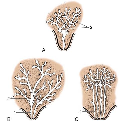
Figure 10-31 Developing duct systems growing proximally from the tip of the fetal teat. A, Cow, ewe, and goat. B, Mare and sow. C, Bitch and cat (only four primary sprouts are shown). 1, Primary sprout, which gives rise to the lactiferous sinus; 2, secondary and tertiary sprouts, which give rise to the lactiferous ducts.
in a single consolidated complex, the udder. Although the glands of the pig, like those of the dog and cat, remain more distinctly separated, this collective term is sometimes also used in the sow. The number of mammary glands (as well as their duct systems) in the domestic species is shown schematically in Figure 10-32.
The more detailed organization is illustrated by reference to the cow. The glandular tissue is arranged in lobules, each 1 mm or perhaps a little more in diameter and consisting of about 200 alveoli. The milk drains to an intralobular duct that joins others to form a larger interlobular duct (Figure 10-33/2). Interlobular ducts lead in their turn to a system of lactiferous (milk-carrying) ducts that ultimately convey the milk to the relatively large cavity known as the lactiferous sinus (Figure 10-33/2). The lactiferous ducts of successive orders increase in diameter but diminish in number so that only 10 or so enter the sinus. Unlike most ducts, they have alternating narrow and dilated portions; contraction of the muscular wall of the narrow portions holds the milk in the expansions before it is “let down” when the cow suckles or is milked.
The lactiferous sinus extends into the teat and is incompletely divided into gland and teat sinuses (Figure 10-33/3'3") by a constriction. The teat sinus is continued by the papillary duct (Figure 10-33∕√), which opens at the tip of the teat where the orifice is surrounded by a smooth muscle sphincter (Figure 10-33/fi).Corresponding parts can be identified in other species, including those in which each gland contains several small lactiferous sinuses, each served by a separate duct system and each opening independently.
It must be stressed that mammary glands are fully developed and fully functional only at the height of lactation. They are then large and show a predominance of yellow glandular tissue over the paler fibrous stroma. When the dam weans her young, involution sets in and the parenchyma regresses (see Figure 29-48, A); the
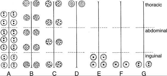
Figure 10-32 Distribution of mammary glands in certain mammals. The dots indicate the number of orifices on the teat. A, Sow. B, Bitch. C, Cat. D, Woman. E, Cow. F, Ewe and she-goat. G, Mare.
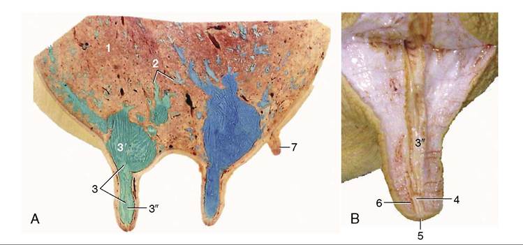
Figure 10-33 A, Sagittal section of udder, showing teat and gland sinuses and lactiferous ducts filled with latex (cranial quarter, green; caudal quarter, blue). B, Section of teat. 1, Parenchyma of gland; 2, lactiferous ducts of various diameters; 3, lactiferous sinus; 3', gland sinus; 3", teat sinus; 4, papillary duct; 5, teat orifice; 6, teat sphincter; 7, supernumerary teat.
connective tissues now form the bulk of the organ. However, the gland never quite reverts to its prelactation size and it grows a little more with each pregnancy.
Mammary buds also form in male embryos and persist to give rise to the rudimentary teats found on the ventral surface of the trunk (carnivores and pig) or on the cranial surface of the scrotum (ruminants). They are less common in horses but occasionally appear beside the prepuce. On the other hand, in certain species, such as rats, the male glands regress completely.