THE PHARYNX AND SOFT PALATE
The pharynx lies behind the mouth and continues into the esophagus. It is a funnel-shaped chamber contained between the base of the skull and the first couple of cervical vertebrae dorsally, the larynx ventrally, and the pterygoid muscles, the mandible, and the dorsal part of the hyoid apparatus laterally.
Because it communicates freely with other cavities in the head, it is rather difficult to form a clear conception of its boundaries and extent; a first impression may be obtained from Figures 3-23 and 4-2. Figure 3-27 illustrates the crossing of the air and food pathways and is a reminder that the pharynx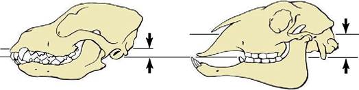
Figure 3-22 The relationships of the articular and occlusal surfaces in the dog and sheep (indicated by the upper and lower arrows, respectively).
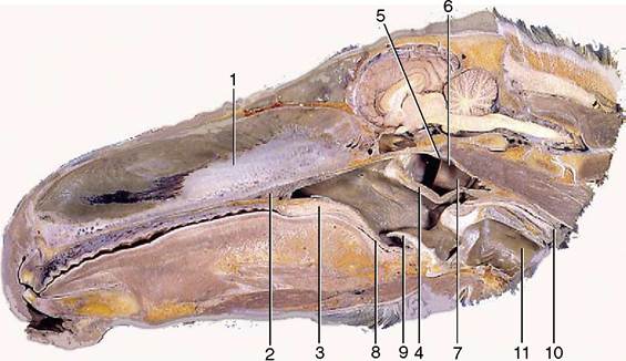
Figure 3-23 Paramedian section through the equine head. 1, Nasal septum; 2, hard palate; 3, soft palate; 4, palatopharyngeal arch; 5, roof of nasopharynx; 6, nasopharynx; 7, entrance to auditory tube; 8, oropharynx; 9, epiglottis; 10, esophagus; 11, trachea.
possesses a respiratory function as well as an alimentary function.
The key to understanding the pharynx is provided by the soft palate, already encountered as the continuation of the hard palate beyond the choanal margin. In repose the soft palate lies on the tongue, but when the animal swallows, the soft palate is raised into a more horizontal position and then more obviously divides the pharynx into dorsal and ventral parts. Two pairs of arches connect the soft palate to adjacent structures. The palatopharyngeal arches pass onto the lateral wall of the pharynx and may be long enough to meet above the entrance to the esophagus (see Figure 3-23).
Together with the free margin of the palate they circumscribe the constriction of the lumen—the intrapharyngeal ostium—that marks the separation of the pharynx into dorsal and ventral compartments. The dorsal compartment is known as the nasopharynx. The more rostral palatoglossal arches pass onto the sides of the tongue at its root; they demarcate the passage from the mouth to the oropharynx (see Figure 3-3). The oropharynx is somewhat arbitrarily divided from the third subdivision, the laryngopharynx, at the level of the epiglottis. The laryngopharynx lies above the larynx and corresponds with this in extent.Functional considerations suggest that the nasopharynx could well be regarded as a part of the nasal cavity. Food does not enter it, it takes no part in the swallowing process, and it serves passively to convey air. The topography of the connection with the nasal cavity varies much among species; a single ductlike communication is present in the dog. In addition to the major connections, the nasopharynx communicates with the cavities of the middle ears through the auditory (Eustachian) tubes. The paired tubal openings are placed on the summits of small pimple-like elevations in the dog.
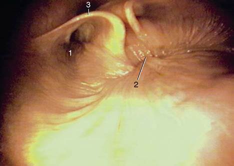
Figure 3-24 Caudal part of nasopharynx (horse). 1, Entrance to auditory tube; 2, closure between the rostral and caudal parts of the nasopharynx (during swallowing); 3, cartilage flange supporting the auditory tube.
Small muscle bundles radiate over the pharyngeal wall from the opening and provide a mechanism for dilating the orifice, thus allowing air to pass to or from the middle ear so that the pressure on the two sides of the eardrum may be equalized (Figure 3-24). Much of the wall of the nasopharynx is reduced to a thin mucosa that finds support by attaching to neighboring structures, mainly the base of the skull and the ventral straight muscles of the head. The mucosa possesses a typical respiratory epithelium and contains numerous mucous glands and much lymphoid tissue, of which some is scattered and some is massed.
The lymphoid masses that form elevations visible to the naked eye are known as the pharyngeal tonsils (adenoids in ourselves) and form part of the ring of lymphoid tissue that guards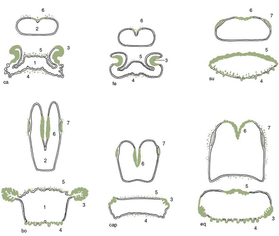
Figure 3-25 Tonsils in the wall of oropharynx and nasopharynx; ca, dog; fe, cat; su, pig; bo, cattle; cap, goat; eq, horse. 1, Oropharynx; 2, nasopharynx; 3, palatine tonsil; 4, lingual tonsil; 5, tonsil of the soft palate; 6, pharyngeal tonsil; 7, tubal tonsil.
the passage from the nose and mouth to the pharynx and beyond (Figure 3-25); like other lymphoid developments they are larger in infancy than later. Excessively enlarged tonsils impair the airflow.
The narrowness of the oropharynx limits the size of the morsels that can be swallowed. Its lateral walls are supported by a fascia and are the site of the palatine tonsils. These are very differently arranged in different species; in some (e.g., the horse) they are diffuse (though raised slightly), whereas in others they constitute a compact mass that may project away from or toward the lumen, as in the ox and dog, respectively (see Figure 3-25). Tonsils that project into the lumen are overlain by flaps of mucosa that partly hide them from inspection through the open mouth (Figure 3-8/8 and Figure 3-26).
The Iaryngopharynx is the largest part of the pharynx. It is wide in front but narrows before joining the esophagus at a boundary that is well defined by a mucosal fold
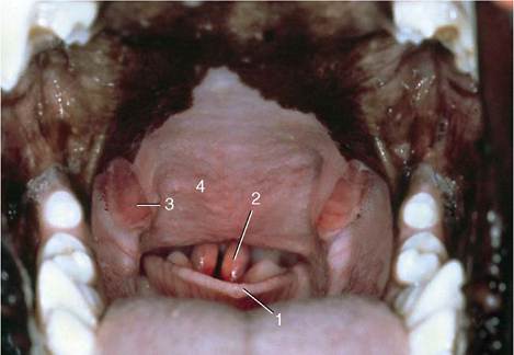
Figure 3-26 View into the oropharynx of a dog. 1, epiglottis; 2, cuneiform process of arytenoid cartilages; 3, palatine tonsils; 4, soft palate.
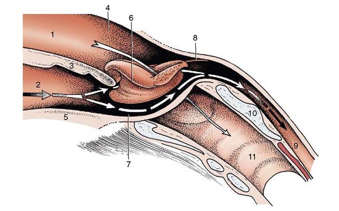
Figure 3-27 Schematic drawing of the pharynx showing its rostral connection with the nasal and oral cavities and caudal connection with the esophagus and larynx.
1, Nasal cavity; 2, oral cavity; 3, soft palate; 4, nasopharynx; 5, root of tongue; 6, larynx (protruding through pharyngeal floor); 7, laryngopharynx (piriform recess); 8, caudal end of palatopharyngeal arch; 9, esophagus; 10, lamina of cricoid cartilage; 11, trachea.in the dog but more difficult to recognize in most other species. At rest, the lumen of the caudal part of the laryngopharynx is closed by the apposition of the lateral walls and roof to the floor. The floor is largely occupied by the entrance to the larynx, which presents the epiglottis, the arytenoid cartilages, and the aryepi- glottic folds. The epiglottis serves as a breakwater to deflect fluids to the side, into gutters (piriform recesses) that run beside the projection of the larynx (Figure 3-27).
Below an external fascia, the greater part of the pharyngeal wall is covered by a set of striated muscles. These fall into three groups—constrictor, dilator, and shortener—although no individual muscle has an action quite so simple as these terms suggest (Figure 3-28). The constrictor muscles arise from certain fixed points conveniently placed to each side and run onto the roof of the pharynx; with their fellows they form a series of arches that enclose the lumen on its lateral and dorsal aspects. For most purposes it is sufficient to recognize rostral, middle, and caudal constrictor muscles, although each may be divided into lesser units. The rostral constrictor arises from the pterygoid region of the skull (pterygopharyngeus) and the aponeurosis of the soft palate (palatopharyngeus) and embraces the pharynx at the level of the palatopharyngeal arch; many fibers take an almost longitudinal course and thus also assist in shortening the pharynx, drawing it onto and over a bolus received from the mouth. The middle constrictor
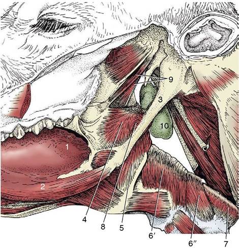
Figure 3-28 Lateral view of the connection of the pharynx with the base of the bovine skull.
1, Root of tongue; 2, styloglossus; 3, stylohyoid; 4, rostral pharyngeal constrictor; 5, middle pharyngeal constrictor; 6, caudal pharyngeal constrictor (6', thyropharyngeus, 6", cricopharyngeus); 7, esophagus; 8, pharyngeal dilator (stylopharyngeus caudalis); 9, tensor and levator veli palatini; 10, medial retropharyngeal lymph node.(hyopharyngeus) arises from neighboring parts of the hyoid bone. The caudal constrictor arises in two parts, from the thyroid (thyropharyngeus) and cricoid (cricopharyngeus) cartilages. When the three constrictors contract in succession, they hurry the bolus distally into the esophagus. The dilator muscle (stylopharyngeus caudalis) also arises from the hyoid apparatus but runs more transversely to fan out in the pharyngeal wall; when active it widens the rostral part of the pharynx, enabling it to accept the bolus more easily.
A fibroelastic aponeurosis internal to the muscles supports the mucosa. It also provides a median raphe to which many fibers of the paired muscles insert and which, continuing to the skull, serves to fix the whole organ in position. The mucous membrane of the oral and laryngeal parts of the pharynx is covered by a stratified squamous epithelium and possesses many small salivary glands that provide additional lubrication to the passage of food.
The soft palate (velum palatinum) is bounded by a respiratory mucosa on its dorsal surface and an oral mucosa ventrally. It is braced by a stout aponeurosis below the dorsal mucosa; the part ventral to the aponeurosis mainly consists of close-packed salivary glands, interrupted toward the midline by the longitudinally disposed palatinus muscle, which shortens the palate. Two small muscles that arise from the muscular process of the temporal bone insert into the lateral part of the aponeurosis after following slightly different courses. As their names indicate, the muscles, the tensor veli palatini and the levator veli palatini, tense the soft palate by exerting lateral traction and raise the soft palate, respectively. The mucous membrane of the pharynx and soft palate and the muscles, except the tensor, which is supplied by the mandibular nerve, obtain their innervation from a plexus to which the vagus nerve makes the chief contribution and the glossopharyngeal nerve a minor contribution.