THE MUSCLES OF THE HEAD AND VENTRAL PART OF THE NECK
The principal groups into which the muscles of the head may be divided are given in Table 2-2, which draws
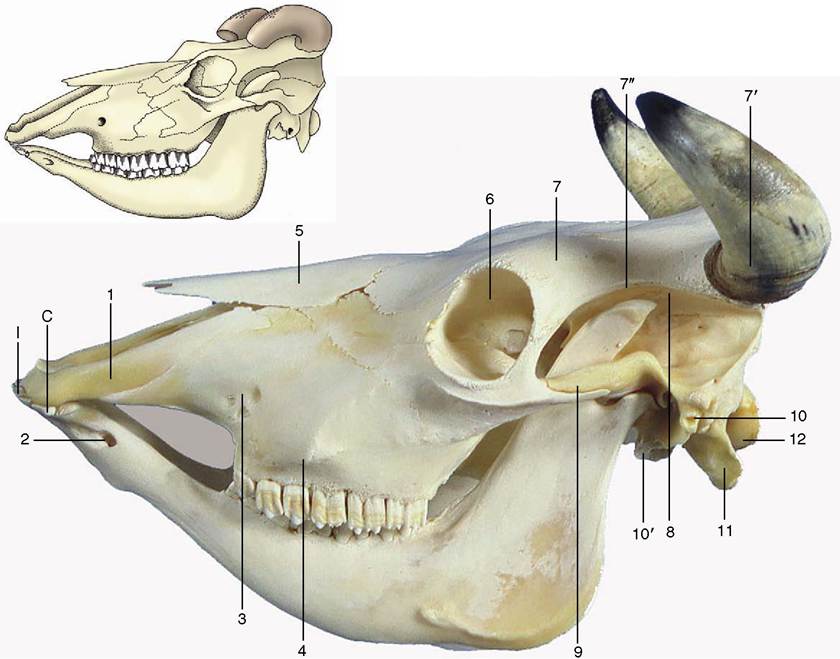
Figure 2-38 Bovine skull with mandible.
1, Incisive bone; 2, mental foramen; 3, infraorbital foramen; 4, facial tuberosity; 5, nasal bone; 6, orbit; 7, frontal bone; 7’, horn surrounding cornual process of frontal bone; 7", temporal line; 8, temporal fossa; 9, zygomatic arch; 10, external acoustic meatus; 10', tympanic bulla; 11, paracondylar process; 12, occipital condyle; I, incisors; C, canine tooth, incorporated in the row of incisors.attention to the correspondence between embryological origin, innervation, and function. The functional associations are so well defined and specific that it is both more convenient and more profitable to refer treatment of most groups to other chapters, where they are considered together with related organs.
The first four groups take origin in the unsplit mesoderm, which covers the lateral and ventral walls of the pharynx and condenses to form the cores of the pharyngeal arches.
In lower vertebrates the muscles equivalent to the last two groups in Table 2-2 are known to develop from somites that appear to each side of the hindbrain, some rostral to the otocyst, the primordium of the inner ear, and the others caudal to it. A similar origin may be assumed in mammals, although the evidence for the formation of these somites is unconvincing at the least. They are of course somatic muscles with the appropriate type of innervation.
The Trigeminal Musculature
The muscles of mastication constitute the greater part of the musculature supplied by the mandibular division of the trigeminal nerve, the motor nerve to the first pharyngeal arch. They are described in the chapter on the digestive system (p.
113). The same chapter deals with the digastricus—a composite muscle to which the mandibular field makes a contribution; the mylohyoideus (p. 105), which slings the tongue between the lower jaws; and one (tensor veli palatini) of the muscles of the soft palate (p. 119). The tensor tympani is considered with the middle ear (p. 346).The Facial Musculature
The musculature supplied by the facial nerve, the nerve of the second pharyngeal arch, is resolvable into two divisions. The superficial division comprises the cutaneous muscle of the head and neck in addition to many small units that control the posture of the lips, cheeks,
| Table 2-2 Source and Innervation of the Principal Muscle Groups of the Head | ||
| Muscle Group | Source | Innervation |
| Masticatory musculature Mimetic musculature Pharyngeal and palatine musculature Laryngeal musculature External ocular musculature Lingual musculature | First pharyngeal arch Second pharyngeal arch Third and fourth pharyngeal arches Sixth pharyngeal arch Hypothetical preotic somites Hypothetical postotic somites | Mandibular division of trigeminal nerve (V3) Facial nerve (VII) Glossopharyngeal (IX) and vagus (X) nerves Vagus nerve (X) Oculomotor (III), trochlear (IV), and abducent (VI) nerves Hypoglossal nerve (XII) |
nostrils, eyelids, and external ears. The deep division is rather scattered but includes some muscles associated with the hyoid apparatus, a contribution to the digastricus (p. 114), and the stapedius (p. 348) of the middle ear.
The Superficial Division. The muscles of this division are conjectured to have their source in an ancestral deep sphincter muscle of the neck, which may be envisaged as arranged in three incomplete overlapping layers. The outermost layer, consisting of transversely disposed fascicles, is reduced to insignificance or is entirely lacking in domestic mammals.
A remnant (sphincter colli) survives in the dog. A more substantial portion of the middle layer commonly persists in the form of a sheet of longitudinally disposed fibers that covers the ventral part of the face and extends onto the neck, even reaching the nape in the dog. It is known as the platysma. Detached slips are believed to provide the small muscles that attach to the caudal aspect of the external ear.The third and deepest layer is again transverse. Although little of it remains in sheet form, it is believed to be the origin of the many discrete muscles of the mammalian face. These are extremely variable among species, but fortunately, few units, and even fewer differences, require detailed notice. Because of their effect on the appearance of the face, they are collectively known as the muscles of facial expression or mimetic musculature.
The principal muscles of the lips and cheeks are the buccinator, orbicularis oris, caninus, levator nasolabialis, levator labii superioris, and depressor labii inferioris (Figures 2-39 and 11-6). The buccinator (Figure 2—39/4) passes between the margins of the upper and lower jaws and is partly covered by the masseter. It forms the basis of the cheek and acts in opposition to the tongue, preventing food from collecting in the vestibule by returning it to the central cavity of the mouth. The buccal salivary glands are scattered among its fascicles, and discharge of their secretion into the mouth may be assisted by contraction of the muscle. The orbicularis oris (Figure 2—39/1) surrounds the mouth opening, where it is closely attached to the skin and mucosa of the lips. It closes the opening of the mouth by pursing the lips and is important in sucking. The caninus (Figure 2—39/2) arises ventral to the infraorbital foramen and radiates into the wing of the nostril and the upper lip. It dilates the nostril and elevates the corner of the mouth in the snarling gesture, especially in the dog. The levator nasolabialis (Figure 2-39/5) arises over the dorsum of the nose and inserts partly on the wing of the nostril and partly into the lateral part of the upper lip.
It is able to dilate the nostril and to elevate and retract the upper lip. The medial part of the upper lip is elevated by the separate levator labii superioris (Figure 2-39/6). This muscle arises on the lateral aspect of the face and runs dorsorostrally to form with its fellow a common tendon that descends into the lip between the nostrils. A special depressor labii inferioris is present in the lower lip of certain species (excluding the dog and cat). It appears to be a detachment from the buccinator muscle. Other muscles associated with the lips and nostrils do not merit specific mention, although some are identified in various illustrations.The muscles of the eyelids include one, the levator palpebrae superioris, that is clearly foreign to the facial group because it arises within the orbit and is supplied by the oculomotor nerve. It is described on page 342. The muscles of the lids that are supplied by the facial nerve include a sphincter—the orbicularis oculi (Figure 2-39/7)—that surrounds the palpebral fissure, the opening between the lids. It is anchored at the medial and lateral commissures and therefore narrows the opening to a horizontal slit when it contracts. Other muscles are present to raise the upper (levator anguli oculi) lid and to depress the lower (malaris) lid, enlarging the eye opening.
The muscles of the external ear are especially numerous but of little account individually. A caudal group
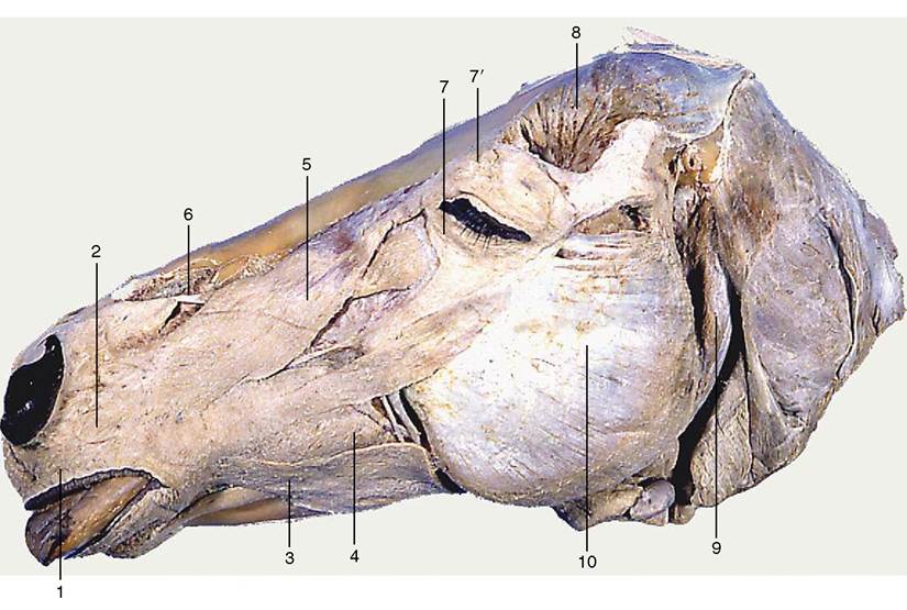
Figure 2-39 Superficial muscles of the equine head. The cutaneous muscle has been removed. 1, Orbicularis oris; 2, caninus; 3, depressor labii inferioris; 4, buccinator; 5, levator nasolabialis; 6, levator labii superioris; 7, orbicularis oculi; 7, levator anguli oculi medialis; 8, temporalis; 9, occipitomandibular part of digastricus; 10, masseter.
has already been mentioned. Others converge on the auricle—the skin-covered cartilaginous ear “trumpet”— from medial, rostral, and lateral directions; they lie between the skin and the temporalis muscle and skull and form a thin, incomplete sheet that includes a (scu- tiform) cartilage plate. The scattered origins and precisely located insertions provide for displacement and rotation of the ear in all directions.
One, the parotido- auricularis, is of somewhat greater importance because it is encountered in the operation for drainage of infections of the external ear of the dog (p. 399). As its name suggests, it arises from the fascia over the parotid gland and approaches the auricle from the ventrolateral direction.Besides the individual functions mentioned or implied in the preceding paragraphs, these muscles have a collective function in communication, mainly within the species but also between species. Human observers can intuitively, or as the result of experience, interpret many facial gestures of animals: one need only recall the hangdog expression of submission, the evident threat conveyed by snarling or laying back the ears, or the quizzical look a dog may adopt. The analysis of the more subtle expressions in terms of specific muscle activity is not yet possible for domestic species.
Paralysis of these muscles is not uncommon after damage to the facial nerve. Since different groups are supplied by branches of the nerve that arise at different levels, the particular pattern of distortions can be a valuable pointer to the location of the nerve lesion (p. 318).
The Deep Division. The muscles attaching to the hyoid apparatus are a rather heterogeneous assemblage. Certain small units are supplied by the facial nerve and elevate the hyoid, in consequence drawing the tongue backward. Although it cannot be denied that these activities have significance in swallowing, the muscles do not appear to merit description. The digastricus, in part derived from the facial musculature, is described on page 114; the stapedius of the middle ear is described on page 348.
The Muscles of the Pharynx and Soft Palate These are considered beginning on page 116.
The Muscles of the Larynx
These are considered beginning on page 153.
The External Muscles of the Eyeball
These are considered beginning on page 341.
The Muscles of the Tongue
These are considered beginning on page 104.
The Muscles of the Ventral Part of the Neck
The neck connects the head with the trunk and is usually distinguished by its relatively slender construction, although this is hardly true of the pig. It has a generally cylindrical form in the dog and cat but is quite obviously compressed from side to side in the larger animals, in which it deepens considerably toward its junction with the thorax (Figure 2-40). The core structures of the neck—the cervical vertebrae and the muscles closely applied to them—were described with the trunk (p. 47). Certain superficial muscles are considered under the heading of girdle muscles of the forelimb (p. 82). The present section is therefore concerned only with the
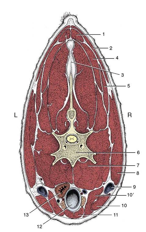
Figure 2-40 Transverse section of the bovine neck. 1, Rhomboideus; 2, trapezius; 3, nuchal ligament; 4, splenius; 5, Omotransversarius; 6, vertebra; 7, longus colli; 8, brachiocephalicus; 9, external jugular vein in jugular groove; 10, 10', sternocephalicus, mandibular, and mastoid parts; 11, combined sternohyoideus and sternothyroideus; 12, trachea; 13, esophagus (ventral to it, nerves, blood vessels, and thymus).
ventral part of the neck, a region of considerable clinical importance on account of the numerous visceral, vascular, and nervous structures that traverse it en route between the head and thorax.
These structures, with the important exception of the external jugular veins (Figure 2-40/9), occupy a central visceral space. The roof of this space is provided by the muscles immediately ventral to the vertebrae, namely the longus colli, longus capitis, rectus capitis ventralis, and scalenus (p. 48). The side and ventral walls blend together and are provided by thinner muscles disposed with a sagittal course and joined by stout fasciae.
The cervical part of the cutaneous muscle (m. cutaneous colli) is unimportant in the dog and cat. It is much better developed in the ungulates, in which it radiates from a stout origin on the manubrium of the sternum; it thins as it passes cranially and laterally and eventually fades away. In the horse, the cutaneous muscle provides a relatively thick cover to the caudal third or so of the jugular groove.
The straplike sternocephalicus (Figure 2-41/2) is the most ventral of the other muscles. It also arises from the manubrium and is first pressed against its fellow. As it ascends the neck, however, it diverges laterally toward its insertion, which varies among species but includes one or the other (or both) of the angle of the mandible and the mastoid process of the skull. The divergence of the right and left muscles exposes the upper part of the trachea to palpation through the skin, although a very thin layer of deeper muscle still intervenes. The sterno- cephalicus is supplied by the ventral branch of the accessory nerve. Unilateral contraction draws the head and neck to that side. Bilateral contraction flexes the head and neck ventrally. In species with a mandibular insertion the sternocephalicus may assist in opening the mouth.
The sternocephalicus forms the ventral border of the jugular groove. The dorsal border of the groove is furnished by the brachiocephalicus, described more fully elsewhere (p. 83). The groove is often visible in life, particularly toward the upper part of the neck. It accommodates the external jugular vein (Figure 2-42).
The deeper muscles constitute an infrahyoid group closely integrated in arrangement and function. They provide an incomplete cover to the lateral and ventral aspects of the trachea and insert, directly or indirectly, on the hyoid apparatus, which they stabilize and retract toward the thorax during swallowing. The obvious members of the group are the sternothyroideus, sternohyoideus, and omohyoideus; the thyrohyoideus on the lateral aspect of the larynx may be regarded as a detached member. The nerve supply is mainly, although possibly not entirely, from the first and second cervical
nerves.
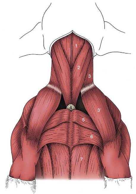
Figure 2-41 Ventral muscles of the canine neck and thorax. 1, Combined sternohyoideus and sternothyroideus; 2, sterno- cephalicus; 3, 3', brachiocephalicus: cleidocervicalis, cleido- brachialis; 4, manubrium of sternum; 5, pectoralis descendens; 6, pectoralis transversus; 7, pectoralis profundus.
The sternothyroideus and sternohyoideus are very thin ribbonlike muscles that take a common origin from the manubrium of the sternum. The caudal parts of the right and left muscles are not always distinctly divided, and in the middle of the neck they may share a common intermediate tendon from which three or four slips diverge cranially. The sternothyroideus inclines laterally to terminate on the lateral aspect of the thyroid cartilage. The sternohyoideus, not always separable from its fellow, passes beside the midline to insert on the basihyoid.
The omohyoideus, lacking in carnivores, is also thin and straplike. Its absence is compensated by the relative enlargement of the other muscles. In the horse it arises from the subscapular fascia, and in the ruminants from the deep fascia of the neck; thereafter it edges medially to join the lateral margin of the sternohyoideus beside which it inserts. In the horse it provides a floor to the
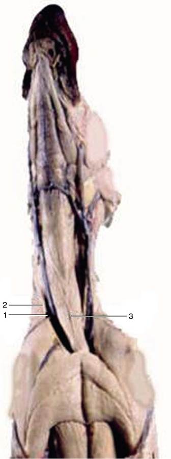
Figure 2-42 Plastination specimen of the ventral part of the neck of a dog. Notice the external jugular vein (1) in the groove formed by the brachiocephalic muscle (2) dorsally, and the sternocephalic muscle (3) ventrally.
caudal part of the jugular groove, separating the vein from the structures within the visceral space.