THE PARANASAL SINUSES
The extensive sinus system possesses considerable clinical interest as it is susceptible to infection that may spread from the nose or from an alveolar abscess. It also provides a means of access to the unerupted portions of the caudal cheek teeth (Figure 18-13).
On each side there are frontal, caudal maxillary, and rostral maxillary sinuses of importance and sphenopalatine and ethmoidal spaces of less account. The layout is complicated and, in one important respect, unique (among domestic species); the frontal sinus communicates with the nasal cavity indirectly via the caudal maxillary sinus.
The frontal sinus occupies the dorsal part of the skull medial to the orbit. It overlaps both cranial and nasal cavities, and because it also occupies the closed part of the dorsal concha, it is more correctly known as the conchofrontal sinus. Its extent is shown in Figure 18-14/7,7'. From this it will be seen that the interior of the frontal part is incompletely divided by several bony lamellae. The floor of this part is molded over the ethmoidal labyrinth, and rostrolateral to these areas of unevenness, it displays the large oval communication (frontomaxillary opening) with the caudal maxillary sinus. The opening normally allows easy natural drainage. A window may be opened, usually by trephination, in the roof of the frontal sinus to allow for irrigation or for removal of a molar by repulsion, when a punch introduced through the frontomaxillary opening is brought to bear on the appropriate alveolus. Such a window also allows introduction of a fiberoptic endoscope to inspect the interior of this large sinus.
The two maxillary sinuses together occupy a large part of the upper jaw, where they have a critically important relationship to the embedded portions of the caudal cheek teeth. They share a slitlike communication (nasomaxillary opening) with the middle meatus of the nasal cavity but are otherwise completely divided by an oblique septum.
This is variable in position but most commonly located about 5 cm caudal to the rostral end of the facial crest. The ventral part of each sinus is also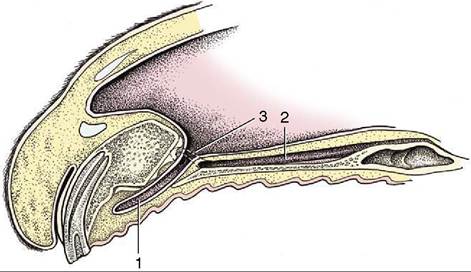
Figure 18-12 Paramedian section of the rostral end of the nose. 1, Incisive duct; 2, vomeronasal organ; 3, opening of the incisive duct into the nasal cavity and opening of the vomeronasal organ into the incisive duct.
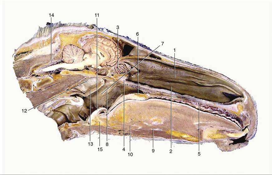
Figure 18-11 Median section of the head; most of the nasal septum has been removed. 1, Dorsal nasal concha; 2, ventral nasal concha; 3, ethmoidal conchae; 4, right choana; 5, hard palate with prominent ridges (rugae); 6, soft palate; 7, nasopharynx; 8, pharyngeal opening of auditory tube; 9, geniohyoideus; 10, genioglossus; 11, epiglottis; 12, medial wall of guttural pouch; 13, pharyngeal muscles; 14, cerebellomedullary cistern; 15, basihyoid.
divided into medial and lateral spaces by an upright longitudinal plate supporting the infraorbital canal and fused in young animals to the alveoli containing the roots and unerupted portions of the cheek teeth. The medial part of the caudal sinus continues into the irregular sphenopalatine sinus. The corresponding part of the rostral sinus extends into the ventral concha.
It is impossible to define the exact extent and projections of the maxillary sinuses, which enlarge considerably after birth as the teeth are extruded (Figure 18-15). Their relationship to the teeth is also affected by the forward migration of the teeth as they develop and come into wear. As Figure 18-15 shows, the relationship is confined to the last premolar and first molar tooth in the newborn foal; it later extends to involve the last four teeth but finally retains contact only with the three molars. There is much variation, and attention to the varying inclination of the embedded parts of different teeth is required.
The surface projection of the maxillary sinuses is considerably larger than the safe surgical field. The latter is determined by several factors, not least the routes followed by the very vulnerable nasolacrimal duct and infraorbital nerve. The potential operating area is defined by the following boundaries: (1) the vertical line tangential to the rostral limit of the orbit; (2) the facial crest; (3) the oblique line joining the rostral limit of the crest to the infraorbital foramen; and (4) the line parallel to the facial crest that intersects the infraorbital foramen. Entry to the sinus may be required either to effect drainage (because the natural route, the nasomaxillary opening, is placed
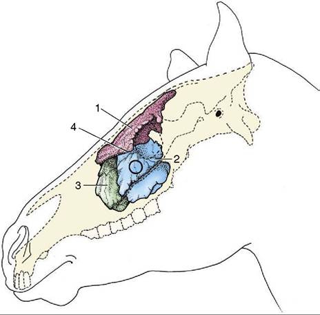
Figure 18-13 Topography of the conchofrontal and maxillary sinuses, which are filled with casting material. The circle indicates where the caudal maxillary sinus can be trephined. 1, Conchofrontal sinus; 2, caudal maxillary sinus; 3, rostral maxillary sinus; 4, position of frontomaxillary opening between 1 and 2.
high in the wall) or to give access to certain teeth.