THE HOOF
The distal extremity of the limb is protected by the hoof, which is formed by epithelial keratinization over a greatly modified dermis; this is continuous with the common dermis of the skin at the coronet (the term applied to the junction between skin and hoof).
The hoof is conveniently divided into wall, periople, sole, and frog; the last is an integral part of the hoof capsule, although homologous with the digital pad of other species (see Figure 10-18).The wall is the part of the hoof visible in the standing animal (see Figure 10-20). It is highest at its dorsal segment (toe) and decreases in height over the sides (quarters) until it is reflected on itself, forming the rounded heels at the back of the hoof. The inflected parts continue forward for a short distance as the bars that are visible beside the frog when the hoof is raised (Figure 23-30/1'"). The angle that the toe makes with the ground is about 50° in the forelimb and slightly more in the hindlimb; the quarters descend toward the ground more steeply, especially on the medial side. The wall is thickest at the toe and gradually thins toward the bars, which is an important point for farriers to bear in mind when rasping or driving nails.
The wall grows from the epithelium covering the coronary dermis (Figure 23-31/2) (which almost surrounds the digit at the coronet). It consists of horn tubules embedded in less structured intertubular horn and slides over the dermis covering the coffin bone and hoof cartilages to be worn away by contact with the ground. The greater part forms the generally pigmented stratum medium. The deeper, nonpigmented stratum internum comprises about 600 (homy) laminae that interdigitate with the sensitive laminae of the underlying laminar dermis (Figure 23-31/5). Trauma affecting the coronary dermis causes horn defects that descend with the wall, reaching the ground in about 8 months (a rate of growth of less than 1 cm per month).
The periople contributes the stratum externum of the wall (Figure 23-31/6,6'). It consists of a band of soft, rubbery horn a few millimeters thick near the coronet but dries to a thin glossy layer distally. The band widens toward the palmar aspect where it covers the bulbs of the heels and blends with the base of the frog. The periople, which also consists of an admixture of tubular and intertubular horn, is produced over the narrow perioplic dermis (Figure 23-31/1) directly proximal to the coronary dermis.
The sole fills the space between the wall and frog and forms most of the undersurface of the hoof (see Figure 23-33). It is slightly concave so that only the distal edge of the wall and the frog make contact on firm ground. The parts between the bars and quarters, known as the angles of the sole, are the seat of “corns,” blood-soaked flecks resulting from trauma to the underlying dermis. Sole horn, though softer than that of the wall, again consists of an admixture of tubules and intertubular horn; it tends to become spongy and to flake in animals required to stand on soiled bedding.
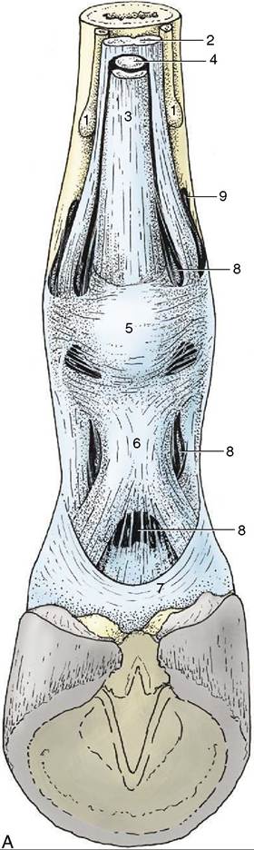
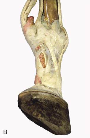
Figure 23-29 A, Annular ligaments of the digit. 1, Splint bones; 2, interosseus; 3, superficial digital flexor; 4, deep digital flexor; 5, palmar annular ligament; 6, proximal digital annular ligament; 7, distal digital annular ligament; 8, digital sheath; 9, palmar pouch of fetlock joint. B, Digital sheath injected with pink, fetlock joint with red latex.
The junction between the sole and the wall is known as the white line (zona alba; Figure 23-30 and Figure 23-32, A). It includes some of the nonpigmented stratum medium of the wall, the distal ends of the horny laminae (stratum internum), and, between these, pigmented horn produced over the terminal papillae of the laminar dermis (these project distally, level with the dermal papillae above the sole) (Figure 23-33/3).
The startlingly white streak within the broad, so-called white line is provided by “cap horn” produced over the distal third of the dermal laminae. The internal rim of the white line is where farriers place nails when shoeing; the nails pass obliquely through the wall to emerge a few centimeters above the shoe, where they are cut and clinched (see Figure 23-30).The wedge-shaped frog (cuneus ungulae) projects into the sole from behind. Its wide base closes the gap between the heels, where it furnishes the palmar part of the hoof (Figure 23-30/4) and spreads upward to end in thickenings—the bulbs of the heels—that overhang the heels of the wall. Its external surface is marked by a central groove to which corresponds an internal spine (frog-stay) that juts proximally into the digital cushion (see further on). The frog is separated from the bars and the sole by deep (paracuneal) grooves (Figure 23-30/6) that accentuate its medial and lateral borders; the
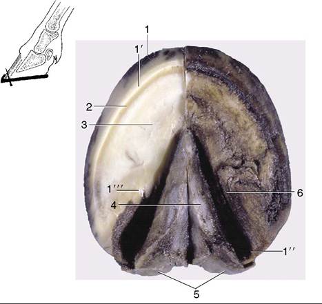
Figure 23-30 Ground surface of the hoof. The inset shows the direction of hoof nails started at the white line. 1, Wall; 1, unpigmented part of wall; 1", heel; T", bar; 2, white line (union of wall and sole); 3, sole; 4, frog; 5, bulbs of the heels; 6, paracuneal groove.
grooves are convenient for the application of hoof testers (large “pincers” used to detect soreness in deeper structures). The projection of these structures is shown in Figure 23-33. In horses stood on damp bedding, the grooves are often the site of “thrush,” a foul-smelling infection that may spread to deeper, sensitive tissues.
The frog horn is tubular and fairly soft and elastic, being kept pliable by the fatty secretion of glands in the underlying digital cushion. Though horses can be shod with the “frog off the ground” (as were city draft horses formerly), a sound hoof requires the frog pressure that is obtained through ground contact.
The dermis deep to the hoof capsule can be divided into five parts: perioplic, coronary, and laminar dermis and those of the sole and frog that are associated with the like-named segments of the hoof. Both the coronary and laminar dermis are associated with the wall.
The entire dermis (other than the laminar part) carries papillae that run parallel to each other and to the dorsal surface of the hoof, directed toward the ground. It is richly supplied with vessels and nerves, and an ill-directed farrier’s nail that penetrates the dermis (“quick”) therefore draws blood and causes pain. Because nerves are absent from the hoof capsule, the apposing dermal and epidermal tissues are often designated sensitive and insensitive, respectively.
The subcutis, generally thin, attaches the dermis to such deeper structures as the coffin bone, the hoof cartilages, and the tendons. It is greatly thickened in two places: beneath the coronary dermis (the coronary cushion) and beneath the frog dermis (the digital cushion). These cushions consist of a feltwork of collagenous and elastic fibers interspersed with small islands of fat and cartilage.
The narrow, raised perioplic dermis embraces the digit at the coronet. Studded with short papillae, it widens caudally where it covers the bulbs of the heels (Figure 23-34/7).
The wider elevation of the coronary dermis (Figure 23-34/2) is separated from the perioplic dermis by a shallow groove. Its prominence is due to the rounded underlying coronary cushion. The coronary dermis also follows the coronet, but like the hoof wall, it folds on itself above the heels. It is widely known as the coronary band, although many clinicians interpret this term more widely to include the (external) coronet. The epithelium over most of its surface produces the bulk of the wall; that over the tucked-in distal margin produces most of the unstructured horn of the horny laminae.
The laminar dermis is composed of about 600 sensitive (dermal) laminae that interdigitate with the insensitive (horny) laminae on the deep surface of the wall (Figure 23-32/5-7).
Both sets bear numerous secondary laminae that further secure the wall to the dermis, and ultimately to the coffin bone, while leaving it possible for the horn to slide over the bone.Normally the epithelium covering the sensitive laminae proliferates just sufficiently to allow the wall to slide past. However, it has the capacity to produce additional amounts of (scar-) horn when a defect in the wall must be closed. This potential is utilized even more dramatically in chronic laminitis (founder), a disease in which the normal attachment is loosened and the coffin bone rotates away from the wall; the space in front of the bone becomes filled with irregular horn produced over a new set of sensitive laminae that form near the dorsal surface of the bone.
The dermis of the sole is firmly attached to the undersurface of the coffin bone.
The dermis of the frog lies between the frog and the digital cushion, which occupies the space below the deep flexor tendon and between the cartilages of the hoof (Figure 23-35/6).
The blood supply of the dermis comes from three sets of vessels, all branches of the digital arteries that descend into the hoof to each side of the flexor tendons. Those that arise at the level of the coronet supply the perioplic and coronary dermis, and those that arise opposite the pastern joint supply branches to the digital cushion and the dermis of the caudal aspect of the hoof, including the frog; the vessels of the third set arise from
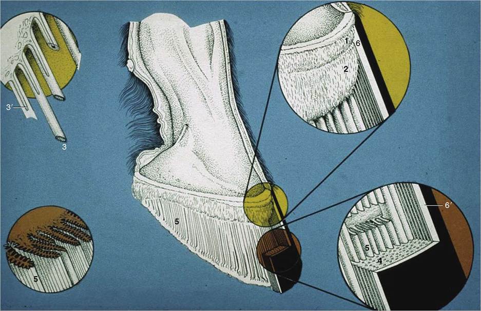
Figure 23-31 The structure of the hoof wall and of the underlying laminar dermis. 1, Perioplic dermis; 2, coronary dermis; 3, horn tubules growing from epithelium over papillae (3) of the coronary dermis (enlarged in left inset); 4, stratum medium of wall consisting of horn tubules embedded in less structured intertubular horn; 5, dermal laminae that interdigitate with the horny laminae of the hoof wall (see also insets to the right); 6, periople; 6', stratum externum of wall (dried periople).
the dorsal and palmar terminal branches (mentioned in connection with the sole foramina of PIII) and go to the laminar and sole dermis. Veins do not accompany the arteries but instead form extensive interconnected networks in the dermis and underlying subcutis, particularly in the coronary band, in the laminar dermis, and under the palmar aspect of the hoof (the coronary, dorsal, and palmar plexuses, respectively). They combine to form medial and lateral digital veins that become satellite to the arteries at the level of the pastern joint.
The hoof is a flexible structure, yielding under pressure on impact with the ground and so dissipating much of the attending concussion. The load that presses on the coffin joint is split between PIII and the navicular bone. The force on PIII is transmitted by the interdigitating laminae to the wall of the hoof, whose distal border is thus a principal weight bearer, especially in horses shod with the frog off the ground. The force retracts the slanted toe while the heels are spread by the distortion of the wall. The force exerted on the navicular bone presses into the yielding “sling” provided by the deep flexor tendon, which in turn compresses the digital cushion and frog (see Figure 23-27). These redirect the force sideways: the cushion presses against the cartilages and the frog presses against the bars and sole, thus assisting the outward movement of the heels (Figure 23-34, C).
The to-and-fro movement of the heels is not obvious to the eye, but as any farrier can verify, it polishes the upper surface of the related parts of the shoe. It is to avoid interfering with this mechanism that farriers do not nail these parts of the shoe to the wall; if this precaution is neglected, the horse develops “contracted heels” and eventually goes lame (Figure 23-34, D).
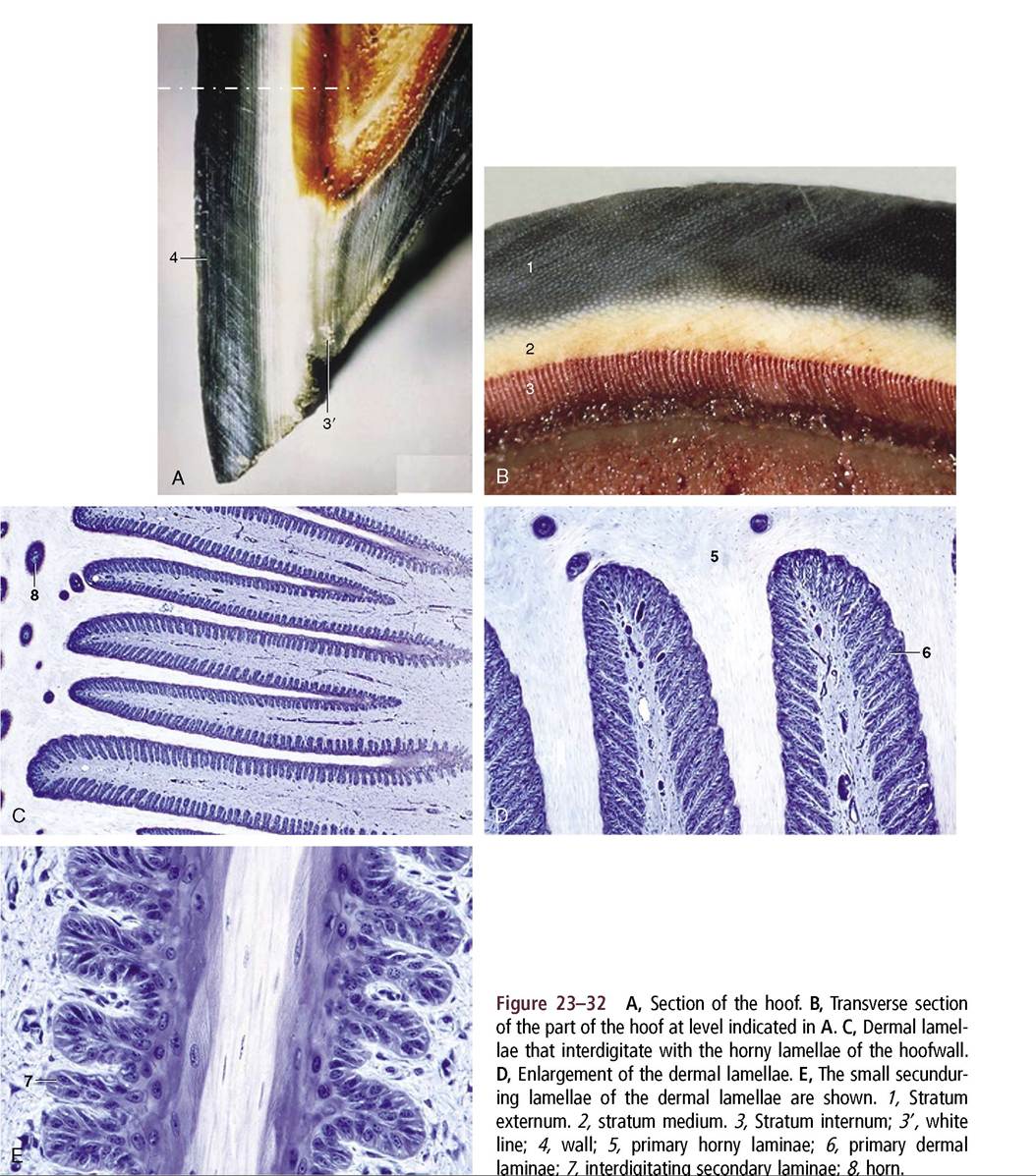
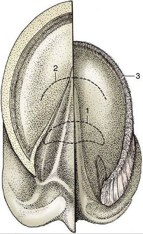
Figure 23-33 Ground surface of the hoof. Half of the hoof has been removed to expose the dermis. 1, Position of navicular bone; 2, position of the insertion of the deep flexor tendon; 3, terminal papillae.
The mechanism explains why the coffin bone is continued caudally by cartilage rather than by bone (Figure 23-21/4). Progressive calcification of the cartilage with subsequent replacement by bone is a common aging process known as “sidebone,” which is yet another cause of lameness.
The movements of the heels have a further benefit, aiding venous return. The dense plexuses on both sides of the cartilages (Figure 23-35/7) are compressed at each step and deliver blood into the valved digital veins. This has been shown experimentally by cannulating a digital vein under local anesthesia; blood is squirted at every step the horse takes. (In other species, contractions of striated muscles within the foot compress the veins and assist the venous return.)
Apart from minor differences in conformation, the forehoofs and hindhoofs are identical (Figure 23-36). In conformity with its larger weight-bearing role, the forehoof is somewhat wider and therefore more rounded in outline than the narrower, more pointed hindhoof (Figure 23-36, C-D). However, the distinction is less than the adjectives suggest, and the provenance—fore or hind—of a single specimen is not always obvious.
When the hoof capsule first forms early in fetal life, it consists of horn that is soft, unpigmented, and of uniform composition. Later, new hard and more structured horn is produced that pushes the soft horn distally, where it becomes a rather misshaped mass covering the entire ground surface of the hoof and (thinly) an adjoining strip of the hoof wall. When exposed to air at term, the soft mass soon dries and sloughs away. The soft mass over the hard horn of the fetal hoof is said to prevent injury to the fetal membranes and birth canal (Figure 23-37).
More on the topic THE HOOF:
- Infectious Foot Rot in Small Ruminants
- Angular Limb Deformities
- Tendinitis
- Endocrinopathic Laminitis
- Heel and Foot Pain
- Physical Examination
- Toxicological Diseases
- Flexural Limb Deformities
- Background Information of Clinical Importance
- Bacterial Diseases