The Placenta Acts as an Endocrine Organ
Besides the essential role of providing nutrients and oxygen for embryonic metabolism, the placenta functions as an endocrine organ. One of the most important functions of the placenta is the production of progesterone.
In primates this function is established early in gestation, and the placenta likely can maintain pregnancy within 2 to 3 weeks after implantation in primates. Placental production of sufficient progesterone to maintain pregnancy occurs later in domestic animals (sheep, day 50 of 150-day gestation; horse, day 70 of 340-day gestation; cat, day 45 of 65-day gestation); in some species the placenta never produces enough progesterone to support pregnancy (cattle, goats, pigs).The production of estrogen, in contrast to that of progesterone, requires interaction between the fetus and the placenta. This interaction has been best described in primates, in particular by the experiments of the Hungarian immigrant to Sweden Dycfaluszy. He and his co-workers found that the primate placenta is unable to produce estrogen from progesterone even though the steroids are only separated by androgens in the steroid biochemical synthetic pathway (see Figure 38-5). The placenta simply does not possess the enzymes necessary for the conversion of progesterone to androgens. Therefore a system has evolved in which the placenta supplies pregnenolone, the immediate precursor of progesterone, to the fetus, and the fetal zone of the adrenal cortex transforms pregnenolone to a C-19 androgen, dehydroepiandrosterone. This is returned to the placenta, which is able to convert dehydroepiandrosterone to an estrogen. In humans the primary estrogen of pregnancy is estriol. Because the fetus is involved in the production of estriol, the well-being of the fetus can be judged by determining estriol concentrations in the plasma of the mother.
The production of estrogen in the mare also involves an interaction between the placenta and fetus (Figure 38-4).
From the work of Pashen and Allen, we know that the fetal gonads replace the fetal adrenals in primates as the key fetal endocrine organ involved in the cooperative synthesis of estrogen. The interstitial cells of the gonads appear to be the interactive cells, with fetal gonads enlarging to a size greater than the maternal gonads during the latter part of gestation. The production of estrogens during pregnancy in other domestic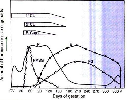
FIGURE 38-4 Summary of the temporal relationships among changes in hormonal concentrations and morphological changes throughout the gestational period of the mare. T CL, Primary corpus Iuteum; 2 CL, secondary corpora Iutea; E, estrogens; E. Cups, endometrial cups; FG, fetal gonads; P, progesterone; PMSG, pregnant mares' serum gonadotropin (equine chorionic gonadotropin). (From Daels PF Hughes JR Stabenfeldt GH: Reproduction in horses. In Cupps PT, editor: Reproduction in domestic animals, ed 4, NewYork, 1991, Academic Press.)
species, occurring relatively late in gestation, may involve the development of placental enzymes that allow progesterone to be metabolized to estrogens without the direct intervention of a fetal endocrine organ. (Fetal cortisol» however, is important for the induction of these placental enzymes, particularly in sheep; see next section.)
The protein hormones that are produced during pregnancy tend to be of placental origin. For example, relaxin is a hormone produced by the placenta in the cat, dog, and horse beginning at about days 20,20, and 70, respectively. Besides its importance for preparing the soft tissues of the pelvic canal for passage of the fetus at birth (see Parturition), relaxin may be important for the support of pregnancy through a synergistic action with progesterone. In exception to the general rule of protein hormone production by the placenta, relaxin is produced by the CL in the pig, cow, and primates during pregnancy, with prepartum release occurring in conjunction with Iuteolysis.
The only CG identified in domestic animals to date is equine CG (eCG, formerly called “pregnant mares serum gonadotropin” by its discoverer, Harold Cole) (see Figure 38-4). The eCG is produced by trophoblast cells that initially form as a band on the chorion (chorionic girdle), detach themselves around day 35 of pregnancy, penetrate the endometrium, and form associations of cells called endometrial cups. The eCG enhances progesterone production by the primary CL of pregnancy and aids in the formation of additional (secondary) CL through the luteinization, or ovulation, of preformed follicles. The essentiality of eCG for pregnancy maintenance is not known, because the primary CL is adequate for maintaining pregnancy.
Placental lactogen is another placental protein hormone. Its production increases in primates as CG secretion wanes during pregnancy. Placental lactogen has been reported in goats and sheep, with secretion increasing during the latter part of gestation. The hormone appears to have both somatotropic and lactogenic effects on the basis of growth hormone-like and prolactin-like properties. In dairy cattle, for example, placental lactogen may be important for mammary gland alveolar development, setting the stage for the next lactation. Another hormone whose production is increased during pregnancy, prolactin, also is important for alveolar development during the prepartum period. Prolactin is not a hormone of placental origin; prolactin increases during the latter part of gestation due to the effect of estrogen on its release from the adenohypophysis.