The Sliding of Actin Along the Myosin Molecule Results in Physical Shortening of the Sarcomere
Figure 6-5 illustrates the sarcomere in the relaxed state and in its shorter, contracted state. The sarcomere is changed from its relaxed state to the shorter, contracted state when Ca2+ ions become available to the sarcomere.
In the presence of Ca2+ ions and a sufficient source of ATP, the actin thin filaments are pulled in parallel along the myosin thick filaments by the repetitive movement of the myosin molecule heads, thus shortening the sarcomere. Because each myofibril is made up of a linear series of repeating and connected sarcomeres, the net result is the physical shortening of the distance between the two ends of the muscle. A more detailed molecular explanation of this sliding filament mechanism of sarcomere shortening is provided in Chapter I as an example of the binding specificity and allosteric interactions of proteins. However, the events can be briefly summarized as follows.At several points along the actin thin filament, there are sites that can bind with the head of the myosin molecule (see
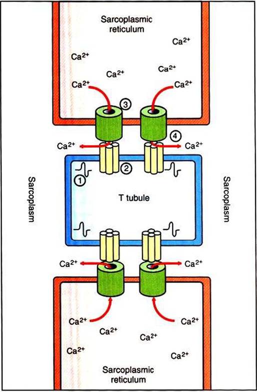
FIGURE 6-4 ■ Relationship between theT tubules (TT) and sarcoplasmic reticulum (SR) during excitation-contraction coupling. 1, Propagation of action potential produces depolarization of theTT membrane. 2, Depolarization induces opening of voltage-gated Ca2* channel aggregates in theTT membrane. 3, Opening of Ca2* release channels on the SR membrane results from mechanical coupling with opening of voltage-gated Ca2* channels on theTT 4, Ca2* is released from the SR into the sarcoplasm, where it can bathe the sarcomeres {not shown) to induce contraction. (Modified from BoronWFz Boulpaep EL: Medical physiology: a cellular and molecular approach, updated edition, Philadelphia, 2005, Saunders.)
Figures 1-4 and 1-5).
In the absence of Ca2+ ions, these sites are either inhibited or covered by the tropomyosin molecules that are normally interwoven within the actin helix. When Ca2+ is present and binds with troponin, a regulatory molecule attached to tropomyosin, the troponin molecule undergoes a configurational change. It is thought that this then causes the tropomyosin molecule to move away from and uncover the myosin-binding site on the actin thin filament, permitting actin-myosin binding. Through a cycle that includes binding with and hydrolysis of ATP, the myosin heads alternately relax and flex while respectively detaching and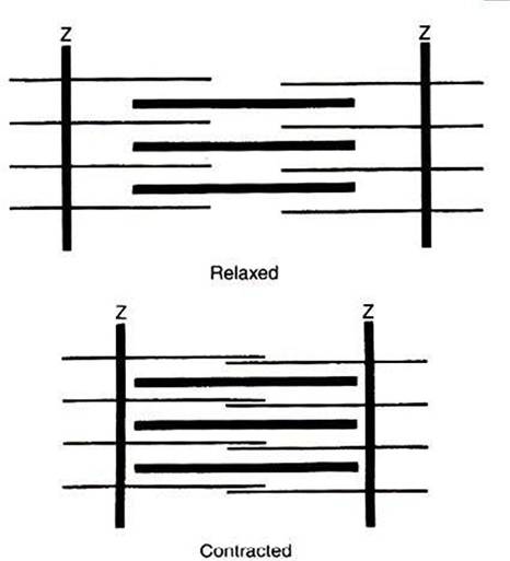
FIGURE 6-5 Sliding of actin along the myosin molecule results in the physical shortening (contraction) of the sarcomere.
attaching to the exposed binding sites on the actin thin filament. This results in the actin thin filaments sliding in parallel along the myosin thick filaments to shorten the sarcomere. In the absence of Ca21, the myosin-binding sites on actin once again become blocked, and sarcomere relaxation results.