The Structural Elements
The cellular basis for the remarkable functioning of the nervous system is the network of interconnected cells known as neurons. Neurons are highly specialized cells in which the membrane properties of excitability and conductivity are extremely well developed.
Rapid, transient changes in electrical potentials travel along neuronal membranes and then are transmitted between neurons within the same circuit through connections known as synapses. A brief description of a highly simplified circuit will serve to explain the manner in which neurons subserve an organism's behavioral reaction to an environmental stimulus (Fig. 8.2). A stimulus, such as change in pressure or temperature, is first detected by a receptor organ (1). Regardless of the type of stimulus, the neuron associated with the receptor (2), normally a sensory neuron in the peripheral nervous system, translates this change into an electrical potential, and the impulse, or action potential, travels the length of the neuron before transmission, via one or more synapses (3), to the next neuron in the circuit. This second neuron (4), located in the central nervous system, receives and in turn transmits the electrical signal to a third neuron within the central nervous system (5). This neuron, the motor neuron, transmits the signal out of the central nervous system to an effector organ in the periphery, normally a muscle (6), resulting in muscle contraction and ultimately movement. Although this description is an oversimplification of any particular neuronal circuitry, it illustrates that the properties of neurons and their organization into anatomic circuits form the underlying basis for nervous system function.The typical neuron consists of the perikaryon, or cell body, containing the nucleus from which extend numerous elongated processes (Fig. 8.3). The processes, which vary considerably in number, length, and form, are of two varieties, the dendrites and the axon.
Dendrites are multiple and highly branched, and they transmit impulses toward the cell body; the axon is always single at its origin at the cell body and conveys impulses away from the cell body. This general morphology underlies four functional regions of the neuron: inputs from other neurons or receptors are received as synapses on dendrites (Fig. 8.3/1); the membrane of the cell body is positioned to support integration of the signals from each dendrite (Fig. 8.3/2); generation of a new electrical signal occurs at the junction of the cell body with the axon; and then transmission of the impulse along the axon occurs, toward synaptic connections with other neurons or with muscle cells (Fig. 8.3/3). The arrangement of the processes results in a wide variety of neuronal morphology (Fig. 8.3D), but superficially permits a simple classification. Most neurons are multipolar in that they possess a number (often a very large number) of branching dendrites that join the perikaryon at scattered points (Fig. 8.3A). Some neurons, predominant in the peripheral nervous system, are bipolar or unipolar. Bipolar neurons possess dendrites that are joined in a common trunk before reaching the perikaryon at a site remote from the origin of the axon (Fig. 8.3B). Neurons with bipolar morphology exist in the retina of the eye and in the olfactory epithelium. The dendrites and axon of a unipolar neuron are directly contiguous along a single process (conventionally known as the axon), and the perikaryon is attached to the axon by a short process (Fig. 8.3C). All sensory neurons in the peripheral nervous system are unipolar in morphology. In both bipolar and unipolar neurons, the integration and generation of the new electrical signal occurs at the confluence of the dendrites rather than at the cell body (Fig. 8.3B and C).The different varieties of neurons have specific distributions that are related to their particular functions. Clearly, a much branched dendritic tree enables a neuron to receive impulses from many sources.
Similarly, a much branched axon makes connection with and stimulates many cells. The first arrangement allows a convergence of impulses from various origins; the second provides for a divergence or diffusion of a message. An axon may establish synaptic connections with the bodies, dendrites, or axons of other neurons, or in the case of the axon of a motor neuron, with the cell membrane of muscle cells. Most neurons establish many synapses: some have many thousands of synaptic sites. Synapses have a variable morphology, but only an elementary description is required here. The synapse is a specialization of both the membrane of the transmitting, or presynaptic, cell, and of the receiving, or postsynaptic, cell. The cell membranes of each are separated by a very narrow gap. An action potential, arriving at the presynaptic terminal membrane, does not jump from cell to cell; instead, it causes the release of a specific chemical transmitter substance that diffuses across the gap. This chemical transmitter is normally stored in vesicles in the presynaptic terminal, awaiting the arrival of the action potential. The arrival of the action potential initiates fusion of the presynaptic vesicle membrane with the cell membrane of the presynaptic terminal, causing release of the chemical transmitter into the synaptic gap. When this substance arrives at the cell membrane of the postsynaptic cell, it binds to receptors embedded in the postsynaptic membrane. These receptors, once bound with the transmitter, produce one of two effects: they can either depolarize or excite the postsynaptic membrane, thus contributing to a new action potential, or they can hyperpolarize the membrane, producing an inhibitory effect by making the generation of an action potential less likely. The existence of both excitatory and inhibitory synapses, sometimes on the same cell, provides a means for a great diversity of response. Many transmitter substances are known; the most common are acetylcholine, glutamate, GABA (gammaaminobutyric acid), noradrenaline, serotonin, and many neuropeptides. This description of synaptic activity as being excitatory or inhibitory is, for brevity's sake, an oversimplification of the complexities of synaptic transmission in the nervous system. Many postsynaptic receptors neither directly contribute to nor inhibit the generation of an action potential but instead make the postsynaptic cell more or less responsive to other transmitters at nearby synapses. This response, termed neuromodulation, permits the fine-tuning of a neuron's response to particular inputs, thus contributing to the subtle nuances of chemical neurotransmission.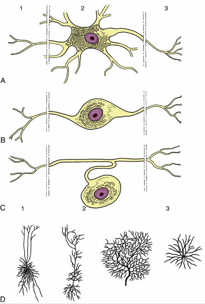
FIG. 8.3 (A-C) Schematic diagrams of (A) multipolar, (B) bipolar, and (C) unipolar neurons. 1, Receptor side (dendrites); 2, cell body (perikaryon); 3, effector side (axon). (D) Drawings of actual neurons.
Neurons are supported by other specialized cells. The supporting cells of the brain and spinal cord, which are much more numerous than neurons, are known as neuroglia and consist of two main types: astrocytes and oligodendrocytes. In brief, astrocytes assist in nutrition of neurons, maintenance of the extracellular environment, and neurotransmission; oligodendrocytes provide axons within the brain and spinal cord with cell membrane sheaths that insulate the axons from their surroundings and speed action potential conductance. The cell membrane sheaths, termed myelin, are formed by the concentric wrapping of oligodendrocyte membrane around the axon (Fig. 8.4).
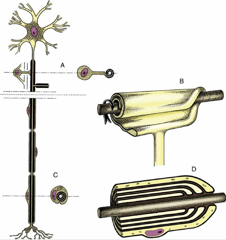
FIG. 8.4 (A) and (B) Neuron with its axon enwrapped within a myelin sheath supplied by oligodendrocytes within the central nervous system. (C) and (D) Once the axon leaves the central nervous system, myelin is provided by Schwann cells in the peripheral nervous system. In both cases, the myelin consists of concentric plasmalemma layers forming an insulating sheath.
Myelin imparts a white color to nerve fibers seen en masse, and groups of myelinated axons in the brain and spinal cord are termed white matter. Within white matter of the brain and spinal cord, axons of common origin, destination, and function tend to be aggregated together into fasciculi or tracts. Most tracts are named by the combination of their origin employed as prefix with their destination employed as suffix. Thus, the spinocerebellar tracts originate in the spinal cord and terminate in the cerebellum; the converse is true for the cerebellospinal tracts. Groups of perikarya, or neuronal cell bodies, in the brain and spinal cord are termed nuclei and, when set off by the whiteness of adjacent fiber bundles, take on a gray or beige color; this effect permits the distinction of gray matter from white matter of the brain and spinal cord.
Within the peripheral nervous system (outside the brain and spinal cord), axons receive insulation similar to that of axons in the brain and cord, but from another type of supporting cell, the Schwann cell (also termed neurolemmocyte; Fig. 8.4). Peripheral axons are grouped together and are protected, supported, and subdivided by connective tissue sheaths and septa, forming the peripheral nerves. The presence of neuronal perikarya in the peripheral nervous system is limited to those of sensory afferent and visceral efferent or autonomic neurons, and they are found in aggregations known as ganglia. Autonomic ganglia on peripheral nerves and sensory ganglia on peripheral nerve roots may form visible swellings; they may also be distinguished by their color and texture, which are darker and firmer than the related nerves or nerve roots.