Bluetongue
Christie E. Mayo • N. James MacLachlan
Definition and Etiology
Bluetongue is an arthropod-transmitted viral disease of domestic and wild ruminants.1,2 Clinical disease is largely restricted to sheep and certain wildlife (notably white-tailed deer, mule deer, pronghorn, bighorn sheep, American bison), but other ruminants, including cattle, may show disease in some circumstances.
Cattle are considered to be especially common reservoir hosts of bluetongue virus (BTV).BTV occurs throughout tropical and temperate regions of the world, where different constellations of BTV serotypes are transmitted by different species of Culicoides midge vectors in relatively distinct global “episystems.”3,4 Whereas infection often occurs year-round in tropical climates, it is distinctly seasonal in temperate areas, occurring only in the late summer and fall months. Bluetongue is usually manifested only in the temperate areas at the upper and lower limits of the global range of the virus. The global distribution of BTV has altered, perhaps as a consequence of the effect of climate change on the Culicoides midges that serve as biological vectors of the virus.5-7 In particular, since 1998 multiple BTV serotypes spread throughout the Mediterranean Basin and, in 2006, additional virus serotypes invaded and spread throughout extensive portions of northern Europe to precipitate an economically devastating epidemic.8 Coincident with this invasion of multiple serotypes of BTV into Europe, at least 10 novel BTV serotypes have also appeared in the southeastern United States.2,9 BTV infection of sheep was detected for the first time in Ontario in 2015, which represents farther northward expansion of the range of BTV in North America.10
Although BTV has been isolated from ruminants or Culicoides midge vectors, or both, from all continents except Antarctica, the international movement of ruminants is highly regulated, and often severely restricted, for fear of importing BTV into bluetongue-free countries or regions.11 The continuing occurrence of disease outbreaks globally, the wide host range, the potential for establishment of endemic infection (as has occurred in extensive portions of Europe that previously were free of the virus), and the similarity of some clinical signs of bluetongue to those of vesicular diseases have all made BTV infection the subject of intense regulatory interest worldwide.
Import regulations promulgated in reaction to the disease have been more of a threat than the disease itself to the livestock industries of some countries.12BTV is the prototype virus in the genus Orbivirus within the family Reoviridae.13 Other closely related viruses in this genus include epizootic hemorrhagic disease of deer and African horse sickness viruses. The genome of BTV includes 10 distinct segments of double-stranded RNA, each of which encodes at least one protein. The serologic classification of BTV into serotypes is based largely on the protein product of just one genomic segment (the L2 gene that encodes the VP2 outer capsid protein), but virulence and other characteristics of individual virus strains may not be related to this protein.14 There is marked genetic variation among field strains of BTV, even viruses of the same serotype and from the same region.15 The genetic diversity and heterogeneity of field strains of BTV arises as a consequence of both genetic shift and drift. Specifically, genetic shift occurs by reassortment of the individual viral gene segments during infections of cells with more than one virus serotype or strain. Individual genes also evolve by genetic drift during alternating replication in the insect and animal hosts of the virus.16
The genetic determinants of BTV virulence are uncertain. At least 30 BTV serotypes are recognized worldwide, but virus strains of the same serotype may have markedly different virulence even to highly susceptible ruminants.1,2,11,17,18 For example, BTV serotype 8 (BTV-8) is present in the Caribbean Basin, where it has not been associated with major outbreaks of disease, whereas the strain of BTV-8 that appeared in northern Europe in 2006 was highly pathogenic and caused significant production loss and mortality in a variety of livestock species, including cattle.3,8,17 Some serotypes of BTV (BTV-25 to BTV-27) infect small ruminants but can have properties quite different from those of the “traditional” serotypes (BTV-1 to BTV-24), notably the capacity for truly persistent infections of ruminants and 1921
for contact transmission 19 21
■ Clinical Signs and Differential Diagnosis Clinical bluetongue disease is manifested in two ways: (1) as reproductive syndromes and (2) as bluetongue per se, a systemic hemorrhagic viral fever that affects multiple organs but especially the upper gastrointestinal tract, skin, and lungs.1,2,17 Sheep most commonly are affected, particularly European fine-wool breeds such as the merino and crossbreeds thereof.
Bluetongue also occurs in cattle, goats, and South American camelids, but less commonly than in sheep and only after infection with especially virulent virus strains.In susceptible sheep, the first clinical signs of bluetongue appear after an incubation period of 3 to 7 days or sometimes longer. These signs include a transient fever (up to 41.1° C [106°F] or higher); edema of the face, lips, muzzle, and ears; excessive salivation; and hyperemia of the oral mucosa (Fig. 32.90). Affected sheep usually have a profuse serous nasal discharge that becomes mucopurulent after a few days, leaving crusts around the nostrils and muzzle. The tongue may be cyanotic (hence the name), although this is a relatively infrequent manifestation. The oral lesions progress to petechial hemorrhages, erosions, and ulcers, which are especially prominent on the dental pad and the commissures of the mouth. The oral lesions can be sufficiently painful that affected animals
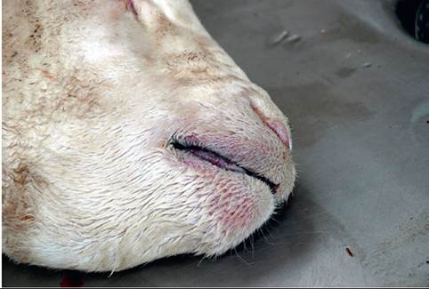
FIG. 32.90 Face of a sheep with severe bluetongue; note submandibular edema and hyperemia and cyanosis of the lips and tongue (bluetongue).
will not eat, and severely affected sheep will stand over water without drinking. Lameness and stiffness caused by coronitis and myopathy can be severe, and the coronary band characteristically shows petechial hemorrhages and hyperemia. Hemorrhages also may be evident in the subcutis of nonhaired skin such as the inguinal region. Lameness may progress to “knee walking” or recumbency. The hooves may slough, and breaks in the wool are common. Diarrhea, with or without blood, can occur. Many sheep become depressed, are unable to rise, and die, but some severely affected sheep make a full recovery. Pulmonary edema is often marked, especially in fatal cases, and some cases appear to livestock owners to be pneumonia. In fact, acute secondary bacterial bronchopneumonia is often superimposed on the characteristic pulmonary edema of bluetongue.
Sudden death, presumably as a consequence of mediator-driven hypovolemic shock or cardiac necrosis and failure, may occur at any time, even in an animal that appears to be recovering. Although the lesions and signs of bluetongue are somewhat characteristic, as is the seasonal occurrence of disease in temperate regions such as much of the United States, the differential diagnosis for sheep showing some of these signs includes sore mouth (contagious ecthyma), FMD, peste des petits ruminants (PPR), and sheeppox.The reproductive and teratogenic effects of BTV in sheep and cattle vary, depending on the virus strain, the host, and other factors. Reproductive effects, including abortions, stillbirths, and weak but live “dummy lamb” births, were profoundly evident when live attenuated vaccine was administered to pregnant ewes in California in the 1950s.22,23 In South Africa, where BTV infection has been recognized as endemic since the first descriptions of the disease before 1900, teratogenic effects have not been commonly linked to the virus despite the use of polyvalent live attenuated vaccines and nearly continuous exposure to the virus.1 However, fetal infections were documented in cattle after these same vaccines were more recently introduced into Europe. Furthermore, it was unquestionably established that these vaccine viruses also circulated naturally after they were introduced into Italy.24 With the notable exception of circulation of live attenuated vaccine viruses, abortion and teratogenic defects are uncommonly associated with natural BTV infection of livestock in endemic areas.25 However, major outbreaks of abortion have occurred after the introduction of virulent BTV into immunologically naive populations of sheep.26-28 Moreover, the strain of BTV-8 that was responsible for the 2006 epidemic in northern Europe crossed the placenta in a substantial proportion of pregnant ruminants, including cattle.8,17 Some authors have questioned whether this remarkable property of this northern European strain of BTV-8 might imply that it was, at some time in its history, propagated in cell culture, as live attenuated vaccine viruses.
Clinical disease is uncommon among cattle infected with most BTV strains, particularly in BTV-endemic areas.1,2,11 However, as evidenced emphatically by the 2006 BTV-8 epidemic in northern Europe, disease certainly can occur in BTV-infected cattle. Severely affected cattle exhibit many of the same signs as do affected sheep. Excessive salivation may be the first clinical sign. Hyperemia and necrosis of the muzzle (“burnt muzzle”) and a patchy dermatitis may also occur, along with necrosis of the skin of the teats. In cattle, depending on which signs are exhibited, the differential diagnosis should include mucosal disease (BVD), malignant catarrhal fever (MCF), vesicular diseases, rinderpest, photosensitization, BPS, and infectious bovine rhinotracheitis. Clinical disease in cattle can also be difficult to distinguish from FMD and vesicular stomatitis, and appropriate regulatory officials should be notified in such outbreaks. Outbreaks in the United States, North Africa, and the Middle East confirm that the related epizootic hemorrhagic disease virus can also cause a bluetongue-like disease syndrome in cattle that also mimics FMD.29
Ocular lesions occur in some BTV-infected ruminants. The syndrome was well characterized during the 2006 BTV-8 epidemic in northern Europe when calves infected transpla- centally in late gestation developed transient corneal opacity (blue-eye) after ingestion of colostrum that contained BTV-8- specific antibody.30 Similarly, non-African ungulates such as European bison at the Berlin Zoological Gardens developed similar ocular lesions after BTV-8 infection.31 “White eye calf” syndrome was described in Oregon in the 1970s and associated with BTV or epizootic hemorrhagic disease virus infection.32 It may be that the lesions result from deposition of immune complexes in the eye, analogous to the immune complex- mediated uveitis that follows immunization of dogs with certain canine adenovirus vaccines.
Laboratory Diagnosis and Immunology
BTV infection in livestock is routinely diagnosed serologically by competitive ELISA that detects antibodies to the core VP7 protein of BTV. The test is highly sensitive and specific and detects antibodies to most if not all BTV serotypes. These antibodies persist for long periods after natural infection, although it is not currently possible to distinguish animals that were naturally infected from those that were immunized with live attenuated vaccine. The presence of antibody indicates only prior exposure to BTV and implies nothing about disease causality. Serotype-specific antibody is currently assessed only with laborious and expensive virus-serum neutralization assays.
Identification of BTV infection in animals is most readily accomplished with a group-specific quantitative PCR assay. Such assays are routinely available in diagnostic laboratories and can identify all known serotypes of BTV. Furthermore, the amount of viral nucleic acid can be quantitated, which can be useful in determining the cause of disease. Acutely affected animals generally have large amounts of BTV nucleic acid in their blood and tissues, which is reflected by low cycle threshold values on the assay. Because ruminants remain positive for BTV nucleic acid for up to 6 months or longer after infection, the detection of either viral nucleic acid by quantitative PCR or of antibody to BTV by competitive ELISA is not proof of disease causality. Serotype-specific PCR assays can be used to serotype the virus present. The availability of these assays has largely obviated the need for virus isolation, which is cumbersome, laborious, and expensive and typically takes several weeks. Virus isolation also requires specialized laboratory facilities because some virus strains require initial propagation in
embryonated chicken eggs before they will grow in cell culture systems. Advances in sequencing technologies have facilitated the characterization of the full BTV genome. Full-length sequencing approaches have helped identify novel serotypes (BTV 25-30), as well as characterize the genetic diversity of the virus. In developing a growing database of full-genome BTV sequences has facilitated the design of conventional and real-time PCR assays. 33
The development of immune tolerance to BTV (in utero infection that results in antibody-negative, virus-positive individuals), as recognized in BVD infections, now is regarded as unimportant in the natural epidemiology of BTV infection, although some controversy persists.17 Fetuses infected with BTV in late gestation are born viremic (virus positive) and, potentially, before they have had the time necessary to develop an immune response. The fact that these animals are born viremic and antibody-negative should not erroneously be interpreted as immunologically tolerant. However, it has been shown that BTV-25 causes persistent infection in goats, although these animals do develop immune responses to the infecting virus that are detected by standard serologic assays. 20
■ Pathophysiology Bluetongue is proposed to be a hemorrhagic viral fever analogous to viral hemorrhagic fevers of humans such as Ebola.17,34 Specifically, BTV replicates within the endothelium and mononuclear phagocytic cells of infected animals. Vascular injury, particularly to small vessels, is responsible for the manifestations of clinical sigms. Release of cytokine and vasoactive mediators from infected cells leads to capillary leakage and, subsequently, the edema and hemorrhage that are characteristic of fulminant cases. Direct virus-induced vascular injury leads to thrombosis and subsequent infarction of tissues, causing ulcers and manifestations such as the characteristic cyanotic (infarcted) tongue in some severely affected sheep. Teratogenic effects result from direct virus- mediated destruction of proliferating progenitor cells within the central nervous system of the developing fetus after transplacental transmission of the virus in pregnant ruminants. Teratogenic defects occur only when the fetus is infected at critical stages of organogenesis, which is before midgestation (approximately 150 days in cattle and 70 days in sheep). Transplacental infection soon after conception can lead to early embryonic death and cause infertility.23
Epidemiology
The development of clinical bluetongue disease is the product of a complex interaction among host, virus strain, vector, and environmental influences.1,2,11 For example, many of the virus strains that circulate in endemic areas such as California are mildly pathogenic for livestock in the region. Epizootics typically occur when a new virus strain or new animals are introduced into a previously stable ecosystem. For example, the 2006 epidemic in northern Europe was the result of the introduction of a highly virulent strain of BTV-8.8 In addition to sheep, this virus caused serious disease in cattle, South American camelids, goats, non-African wild ungulates such as yaks and European bison, and even some carnivore species.31 Environmental alterations that favor the proliferation of midge vectors in a region also promote disease epidemics.
BTV infects wild and domestic animals primarily through the bite of the vector midge of the genus Culicoides. There are more than 1400 Culicoides species globally; however, only a few have been identified as BTV-competent vectors.1-3,5,6,11,35 In temperate regions of the world, Culicoides midges are abundant in midsummer to early fall, and outbreaks of bluetongue in livestock occur during this time when temperatures support both midge activity and BTV replication within the vector. Other routes of virus transmission are possible even during the interseasonal period in temperate regions (winter, spring, and early summer). For example, it has been shown that BTV can be spread to newborn ruminants via the ingestion of infective colostrum.36,37 Lastly, although BTV can be transmitted sexually in infected semen and transplacentally from dam to offspring, the importance of these potential routes of virus transmission remains conjectural; they are unlikely to have a major role.17 Specifically, animals infected in early gestation develop teratogenic brain defects and have cleared infectious virus by the time of birth. Fetuses infected in late gestation can certainly be born viremic and potentially contribute to virus amplification and circulation. Embryo transfer currently is not viewed as a significant risk of BTV transmission if the embryo is washed at least 10 times. In summary, dissemination of BTV by vector Culicoides midges is by far the most common method of transmission in endemic areas; however, some serotypes of BTV (notably BTV-26 and BTV-27, and probably BTV-25) can be spread by direct contact transmission without requirement for the insect vector. 19
In much of North America, the prevalence of bluetongue closely mirrors the distribution and abundance of Culicoides sonorensis, with lower rates in northern climates that are less hospitable to the midge and higher prevalence in California and other warmer southern regions.3,4 However, the vector species in the southeastern United States, where 10 additional serotypes of BTV have appeared, are less defined. Similarly, vector species other than C. sonorensis are important elsewhere in the Americas as they are in different regions of the world. In the absence of competent vector populations, animal-to- animal transmission appears to be incapable of maintaining endemic BTV infection; thus BTV-8 disappeared quickly from most of northern Europe.
It remains uncertain as to how BTV is maintained (overwintered) in temperate areas where infection is highly sea- sonal.2,11 Although viremia can be prolonged in both cattle and sheep, BTV infection is not persistent in domestic ruminants and insufficient to sustain the virus through the several months of the overwintering period (approximately December through June in the northern hemisphere). Infection of vector midges is persistent and lifelong; however, the life span of midges is relatively short, and so it is unlikely that infected individuals could survive the entire overwintering period. Studies in California have confirmed the presence of BTV-infected parous female Culicoides midges in midwinter without concurrent infection of adjacent sentinel cattle, which suggests that long- lived vectors infected in the prior seasonal period of transmission sustain BTV throughout the overwintering period in seasonally 3839
endemic areas. 38,39
The severity of clinical signs in BTV-infected sheep varies by breed. Early in the history of the bluetongue investigation, it was noted that African breeds showed few if any signs of infection, whereas imported European breeds showed fulminant disease. 1,2 More recently, breed differences in immunologic response and susceptibility to disease expression after vaccination with live attenuated BTV vaccine have been reported.40
Necropsy Findings
The lesions of bluetongue are characteristic, particularly in sheep.1,17,41 These include ulcers and hemorrhage throughout the oral cavity, including the dental pad and tongue. These lesions may extend from the esophagus into the forestomachs. Coronitis and subcutaneous hemorrhages and edema, along with edema between the fascial planes of the neck and abdominal musculature, are also common. Other typical findings are pulmonary edema, hemorrhages on the endocardial and epicardial surface of the heart and pulmonary artery, and necrosis of both skeletal and cardiac muscle. Especially characteristic lesions include gelatinous edema around the nuchal ligament (in addition to the other sites previously listed), subintimal hemorrhages in the pulmonary artery, and discrete focal areas of acute myocardial necrosis in the papillary muscle of the left ventricle. Microscopic study reveals cuffing of inflammatory cells around small vessels in the affected skin and gastrointestinal mucosa with pronounced hypertrophy of the endothelium lining affected vessels, acute and focally extensive necrosis within the musculature of the heart and some skeletal muscles, and pulmonary edema.
Treatment, Prevention, and Control
Treatment of bluetongue in affected ruminants is nonspecific and aimed at supportive and nursing care. Animals with severe oral lesions are reluctant to eat or drink. Valuable animals can be fed gruels of alfalfa pellets by stomach tube and can be encouraged to eat soft feeds or green grass. Muscle and coronary band pain may limit mobility; therefore water and shade must be close at hand. Sulfa drugs or other relatively broad-spectrum antimicrobial drugs can be administered in an attempt to prevent or treat secondary bacterial pneumonia. Antiinflammatory medications that include but are not limited to NSAIDs, aspirin, and flunixin are commonly given.
Elimination of C. sonorensis (or other midge vectors) from the environment is usually not practical, but housing sheep indoors during the peak of activity (dusk, early evening) to minimize exposure to biting midges may be beneficial with regard to some vectors. Grazing wet areas such as irrigated pasture only during the heat of the day also may help, as most insects are not active in extreme temperatures. Midges that feed on ivermectin-treated cattle die, but the exchange of virus may occur before the insect's demise. C. sonorensis larvae develop in fine-grained mud with high organic matter content, such as those around farm reservoirs, overflowing watering troughs, and shallow septic systems. Elimination of these breeding grounds along with with larvicidal treatments may help in some situations. Especially valuable animals can be housed in insect-proof enclosures to prevent any contact with vector midges during outbreaks, or they may be treated with repellants to minimize the likelihood of vector attack.
Both inactivated and modified live BTV vaccines are available in some parts of the world, and these logically should be based on the local strains and serotypes.1,2,42,43 Inactivated vaccines are inherently safer than live attenuated ones because the latter have the potential for transmission in nature, reversion to virulence, and the capacity to cross the placenta. There is little cross-protection between BTV serotypes, so animals need to be vaccinated against all serotypes of the virus circulating in a given region. A live attenuated vaccine is available in some western states of the United States for immunization of sheep, but there currently are no vaccines to other endemic serotypes such as BTV-2, BTV-3, BTV-11, BTV-13, and BTV-17.37 Live attenuated vaccines should be given at least 2 weeks before breeding season to avoid teratogenic effects, and vaccination should be done before the seasonal period of virus transmission (late summer and fall) to avoid infection of vectors and so minimize the likelihood of recombination of vaccine and field viruses. Ewes may be vaccinated late in pregnancy, but there is some risk of inducing abortion. Vaccine should not be administered in early or midgestation because of potential teratogenic defects. Vaccinated breeding rams may have some risk of transiently decreased fertility. Vaccinated and naturally infected animals cannot readily be distinguished by current serologic diagnostic tests. New-generation BTV vaccines have been developed, including recombinant vector vaccines, and virus-like particle vaccines. In addition to being very safe, these vaccines also have the potential to allow vaccinated animals to be distinguished from infected animals (so-called DIVA).44
More on the topic Bluetongue:
- CHAPTER MENU
- Insurance, Interstate, and Prepurchase Health Examinations
- Preface to the Third Edition
- Retinal Changes
- Virus-Associated Myopathy
- Specific Diseases of the Digestive System Viral Diseases
- FACTORS DRIVING DISEASE EMERGENCE
- Arthrogryposis
- Consideration of Epidemiology
- Etiologic Diagnoses