General Evaluation of the Patient With Respiratory Disease
Kara M. Lascola • Pamela A. Wilkins
History
As with any disease process, acquisition of an accurate and appropriate history is the first step undertaken in evaluating the patient with a complaint thought to be related to the respiratory tract.
Animals with respiratory disease may have widely varied histories, so a thorough anamnesis is necessary. Age and breed may play a role in the development of respiratory disease, such as congenital defects or neoplastic disease, or in inherited or acquired immunodeficiency syndromes seen in certain breeds. The environment in which the animal is maintained can contribute to the development or severity of respiratory disease. For example, equine recurrent airway obstruction (RAO) and inflammatory airway disease (IAD) may manifest following a change to a new environment. Stressful events, such as weaning or long-distance transport, can predispose both horses and cattle to respiratory disease. It is important to know if certain diseases are either endemic or epidemic where the animal is kept or has recently resided. Any recent traumatic or potentially traumatic event should be noted. A thorough vaccination history should be obtained, as should an accurate history of any treatments or supplements and the patient's response. Presenting Signs or Chief Complaints
Unilateral or bilateral nasal discharge is a common finding or complaint associated with respiratory disease. Respiratory noise at rest or during exercise is commonly associated with abnormalities of the upper airway and may accompany inequalities of airflow present at the nares. Normal animals may periodically cough or sneeze, but an increase in either activity may indicate involvement of the respiratory tract. Exercise intolerance or reduction in athletic performance should prompt evaluation of the respiratory system. Other clinical signs that should encourage the clinician to conduct a thorough evaluation of the respiratory tract include abnormal breathing patterns (tachypnea, hyperpnea, dyspnea), cyanosis, hemoptysis, epistaxis, unusual swellings (facial, pharyngeal, cervical), lymphadenopathy, ataxia or reluctance to move, foul smell to the breath, weight loss, and ventral abdominal, sternal, or limb edema.
Physical Examination
The initial physical examination occurs at some distance from the patient and involves evaluation of the demeanor, posture, mental status, and way of movement of the patient. It is important to note if the patient has an abnormal stance, such as standing with the head and neck extended. If the patient is unwilling to move or stands with elbows abducted, this suggests that pleural pain is present. Ideally the respiratory rate can be determined by observation, as can the respiratory pattern. Although some respiratory diseases are not manifest at rest, important clues can be gained from observation of the patient at rest in many others. The normal resting respiratory rate of an adult horse is 8 to 16 breaths/min, of adult cattle is 15 to 35 breaths/min, and of sheep and goats is 12 to 20 breaths/ min. There is some small abdominal component during the expiratory phase. The normal rate for neonates is up to 60 breaths/min at birth and less than 30 breaths/min by 1 month of age; respiratory rate decreases toward the adult rate with age. High ambient temperature, fever, and excitement can all increase respiratory rate. Normal breathing, which is quiet and apparently effortless, is termed eupnea. The term dyspnea is a breathing pattern that is inferred by the observer to reflect difficulty in breathing; the animal will appear distressed, and the work of breathing is obviously increased, although the actual rate may be within normal limits. Other terms used to describe breathing patterns include tachypnea, characterized by rapid rate and shallow depth or low tidal volume; hyperpnea, with increased frequency and depth of breathing (an example would be during postexercise recovery); and apnea, in which there is no discernible breathing. Two additional terms include hypoventilation and hyperventilation, both of which require a change in arterial carbon dioxide partial pressure as a component of their definitions. Hyperventilation is a pattern that increases alveolar ventilation and causes arterial hypocapnia, whereas hypoventilation alters gas exchange in such a way to cause arterial hypercapnia, or retention of carbon dioxide.
Closer examination can reveal some of the physical manifestations of presenting complaints listed previously. Beginning with the head, the clinician should determine that airflow is even from both nostrils, as differences can indicate either congenital or acquired abnormalities ranging from choanal atresia to the presence of upper airway masses. Abnormal respiratory sounds can sometimes be present at rest and may be heard at the nares. Abnormal breath odors may be detected. The frontal and maxillary sinuses should be percussed; identification of abnormal resonance, usually dullness, may be made easier by performing this with the mouth held open. Palpation of the submandibular regions, larynx, and pharyngeal and cervical regions should be performed to identify any abnormal lymph node enlargement, masses, or areas of muscular atrophy. Both jugular veins should be checked for patency and the presence of any evidence of injection sites or infections that may contribute to abnormal upper airway function by interfering with normal recurrent laryngeal nerve or vagosympathetic trunk function.
Coughing represents a nonspecific irritation of receptors in the airway and can be induced by many mechanisms. It can be, and usually is, a normal protective reflex that allows the animal to clear material from the airway. Cough can be associated with increased mucus production, production of other respiratory secretions, or decreased mucociliary clearance. In older horses cough is often associated with severe RAO, whereas in younger horses cough is more commonly associated with infectious diseases and IAD (see discussion of these disorders later in this chapter). Normal animals should not cough when the larynx or trachea is palpated.
Nasal discharge can be unilateral or bilateral, scant or copious, clear, mucoid, mucopurulent, or even bloody. The nature and character of nasal discharge can provide some information about a possible source of the discharge, but this information should not be overinterpreted.
Horses, for example, have a tendency to swallow excess airway secretions, and the volume of secretions may be underestimated. Whereas unilateral nasal discharge is often suggestive of a source in front of the larynx, bilateral nasal discharge can be of either upper or lower airway origin. Skin depigmentation of the ventral nares or presence of mucoid material in feed or water containers are clues to the presence of nasal discharge. Hemoptysis is the coughing up of blood from the airways or lungs. It is important to determine conclusively that the blood has come from the respiratory system. Epistaxis is defined as blood seen at the nares and often originates in the nasal passages, sinuses, turbinates, nasopharynx, or equine guttural pouches, although the lung can be, and is, a source on occasion, as in exercise-induced pulmonary hemorrhage (EIPH) or following lung biopsy. Bilateral epistaxis generally indicates bleeding caudal to the choanae. Because animals tend to swallow excessive respiratory secretions, bleeding can be occult and may not be seen unless the animal drops its head toward the ground. Significant blood loss can occur in this manner, unseen by owners.
Examination of the oral mucous membranes may reveal cyanosis, which is bluish discoloration of the oral, nasal, or vulvar mucous membranes. Cyanosis does not become apparent until approximately one third of the total normal hemoglobin is deoxygenated; this reflects a profound decrease in oxygen saturation of hemoglobin and is suggestive of severe hypoxemia. Since the total quantity of deoxygenated hemoglobin lends the mucous membranes the bluish color, very anemic patients may lack sufficient deoxygenated hemoglobin to appear blue, which makes appreciation of cyanosis impossible in these patients. One caveat is that all newborns are cyanotic for the first few breaths and only become pink when they have established neonatal, as opposed to fetal, cardiorespiratory circulation and have fully inflated their lungs to allow for gas exchange.
It is important that auscultation of the thorax take place in as quiet an environment as possible. In addition, auscultation of the lung fields should be performed under two breathing conditions, eupnea and hyperpnea, with the latter induced by the use of a rebreathing maneuver (bag). The purpose of this technique is to cause the animal to rebreathe its own expired carbon dioxide. The resultant increase in PaCO2 stimulates deeper and more frequent breathing efforts, making recognition of abnormal lung sounds simpler. The rebreathing bag must be large enough to accommodate two to three times the normal tidal volume of the animal and should be held in such a manner as to prevent the bag from occluding the patient's nostrils. Once the bag is removed, the animal will usually take several very deep breaths, and the examiner should take advantage of these breaths to reexamine areas where suspicious sounds were heard during rebreathing. Coughing and prolonged time of return to baseline respiratory patterns should be noted. This technique is contraindicated in animals demonstrating increased respiratory effort at rest.
Normal breath sounds are those produced by turbulence within the tracheobronchial tree and may vary considerably depending on location within the lung, breathing pattern, and condition of the animal.1 Only airways from the larynx to segmental bronchi contribute to sound generation. Vesicular sounds—the quietest sounds, heard over the middle and diaphragmatic lung regions—correlate best with regional ventilation and mainly represent segmental bronchial sounds; they do not represent airflow in terminal conducting airways and alveoli, which is silent due to the nature of its flow. Bronchial sounds are louder and heard best over the trachea and base of the lung. Common abnormalities found during auscultation include ventral areas of dullness if pleural effusion is significant, dorsal areas of dullness or hyperresonance with pneumothorax, and dorsal harsh lung sounds.
The degree of variation in normal lung sounds is large, and auscultatory findings do not always correlate well with the degree of lung abnormality. That said, abnormal lung sounds are always potentially clinically important. Adventitious lung sounds are divided into short discontinuous sounds called crackles and longer continuous sounds called wheezes, replacing the older terms rales and rhonchi, respectively. Crackles are most commonly generated by sudden pressure equalization when collapsed airway segments open. Although an air-fluid interface is required, crackles do not necessarily imply excessive secretions or pulmonary edema. They are often end-inspiratory and associated with reinflation of atalectatic lung. Crackles may be normal when auscultated in the newborn or over the previously down-side lung of a laterally recumbent neonate. Disease processes that generate crackles include pneumonia, interstitial fibrosis, chronic obstructive lung disease, congestive heart failure, and atelectasis.2
Wheezes commonly represent oscillation of airway walls before complete closing (expiratory) or opening (inspiratory). Intrathoracic airways are usually involved in expiratory wheezes and include the lower trachea and main, lobar, and segmental bronchi. Disappearance of a wheeze after coughing indicates secretory rather than tissue component origin. Disease processes responsible for wheezes include airway stenosis or external compression, airway lumen compromise by a foreign body, purulent material, cyst or neoplasm, bronchoconstriction, and airway wall thickening as occurs in chronic bronchitis. Expiratory wheezes are a hallmark of obstructive lung diseases such as RAO. Crackles and wheezes may be variably present. A final category of adventitious sounds is the “rubbing” or “creaking” sounds generated by sliding or stretching of inflamed pleural surfaces, commonly termed pleural friction rubs.
Percussion of the thorax is performed by methodical tapping over the intercostal spaces of the thorax using a variety of instruments, including plexors, pleximeters, spoons, or fingers. It is of relatively low diagnostic sensitivity but may be useful particularly when thoracic ultrasound is not available. It should be performed when pleural effusion is suspected and in all
ruminants to detect occult pneumonia. Percussion can reveal hyporesonance (dullness) ventrally when pleural effusion is present, reveal hyperresonance dorsally in pneumothorax, and cause some patients with pleurisy to exhibit pleurodynia (pleural pain). Other conditions that can alter resonance of the thorax include diaphragmatic hernia with intrathoracic intestine, pericardial effusion, pulmonary and pleural abscessation, and consolidated lung. It is usually impossible to fully delineate the lung field cranially because of body fat and triceps musculature. There is a distinct region of cardiac dullness for all species on the left side.
Additional Diagnostic Evaluation of the Respiratory Tract
Endoscopy
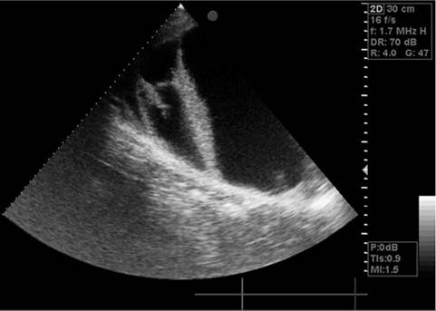
The upper airway can be directly examined with the aid of an endoscope, the only limitations being the size of the patient, the patency of the airway, available means of restraint, and the size of the available equipment. Standard flexible fiberoptic endoscopes allow direct examination of the nasal passages, ethmoid turbinates, nasal maxillary opening of the sinuses, pharynx, guttural pouch openings, larynx, and cranial trachea (Fig. 31.1). Smaller (8 to 10 mm in diameter) endoscopes can be readily introduced into the equine guttural pouches, with the aid of a biopsy instrument, and longer bronchoscopes (200 to 250 cm) are commonly used to examine main stem bronchi and their initial branches in large animals. Smaller species may require the use of a smaller diameter (3 to 5 mm) endoscope or bronchoscope. Small brushes, used for collecting exfoliated cells for cytologic study, and a variety of biopsy instruments can be used for sampling the airway.
Airway endoscopy in the horse has evolved to include dynamic respiratory endoscopy (e.g., high-speed treadmill, overground endoscopy) during exercise to evaluate the dynamic function of the upper respiratory tract.3-5 Objective measurements can be made using videoendoscopy with slow motion or freeze-frame features.3 Treadmill endoscopy is available at specialty practices and typically is performed at high speed (12
![]()
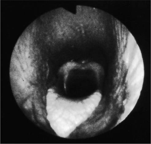
FIG. 31.1 Normal equine larynx. The larynx is directly visualized by endoscopy, with both structure and symmetry evaluated. (Courtesy Dr. Corinne Sweeney, University of Pennsylvania, New Bolton Center, Kennett Square, Penn.)
to 14 m/s) using incremental standardized testing protocols. Overground telemetric endoscopy is performed during ridden exercise in the field with endoscopy equipment that is attached to the horse.4,5 Advantages of overground endoscopy include the ability to evaluate the horse under saddle and in natural working conditions. However, standardizing exercise tests in the field may be more challenging. While resting endoscopy can detect many abnormalities involving the upper airways, dynamic upper airway obstructions that develop during exercise may be missed. In these situations dynamic endoscopy, if available, is the preferred diagnostic tool.5 Sedation or tranquilization will aid many standing endoscopic examinations, but examinations aimed at evaluating pharyngeal and/or laryngeal function are best performed without any form of chemical restraint that might alter function. Most horses will allow standing examination of the upper airway with only physical restraint, such as judicious use of a nose twitch. Introduction of the endoscope into the trachea may elicit coughing. Small ruminants, such as sheep and goats, may require local tracheal administration of 2% lidocaine diluted to a 0.2% to 0.3% solution. If lidocaine is used in small ruminants, it must not reach a toxic dose (~6 mg/ kg). Diluted lidocaine can similarly be used in horses and cattle for evaluation of the distal trachea, main stem bronchi, and larger bronchial tree branches. Horses are more sensitive to tracheal and bronchial stimulation and are more likely to require lidocaine than are cattle.
Guttural pouch diseases and upper airway abnormalities, such as pharyngeal lymphoid hyperplasia, laryngeal hemiplegia, epiglottic entrapment, dorsal displacement of the soft palate, pharyngeal cysts, retropharyngeal masses, and epiglottic deformities, are best diagnosed by endoscopic examination. Direct tracheobronchoscopic examination is useful for the diagnosis of tracheal diseases such as tracheal collapse or trauma as well as for the diagnosis of lower airway abnormalities such as EIPH and mild to moderate equine asthma (IAD). Bronchoscopy may also be useful for evaluating additional lower airway abnormalities, including severe equine asthma (RAO), tumors, or abscesses. The degree and nature of airway secretions accumulating in the trachea can be easily assessed with an endoscope, and scoring systems for tracheal mucus and blood have been developed for the diagnosis of mild to moderate IAD6 and EIPH,7 respectively. Accumulated secretions in the trachea and distal airways may be sampled by aspirating the secretions through small tubing introduced through the biopsy channel of the endoscope. Because the endoscope has passed through the nonsterile upper airway, samples collected without the use of a sterile, double- or triple-lumen guarded tube are best suited for cytologic, not microbiological, evaluation but may be fully compatible with evaluation using molecular diagnostic techniques.8-11 Endoscopy has also been used to help remove foreign bodies from the airway, generally aided by the biopsy instrument.
Endoscopic evaluation of the thoracic cavity (thoracoscopy) can be performed in horses under sedation or general anesthesia12 and has also been described in healthy cattle.13 The procedure can be useful for the evaluation and treatment of conditions such as thoracic neoplasia, pleuropnemonia or other pleural disorders, and thoracic trauma. Thoracoscopic-guided lung biopsies can also be performed in the horse.14 Thoracoscopy is performed using standard laparoscopic equipment, including a rigid endoscope, camera, monitor, light source, and insufflation systems when performed under general anesthesia. The provision of supplemental oxygen is essential, and ventilatory support may become necessary during standing procedures.
Diagnostic Imaging
RADIOGRAPHY. Radiographs are indicated for evaluation of congenital abnormalities, diseases, or trauma involving the respiratory tract or thoracic cavity. Radiographs are frequently performed along with endoscopy or thoracic ultrasound for more thorough evaluation of the upper respiratory tract or lower respiratory tract and thoracic cavity, respectively. Portable equipment needed to perform radiographic evaluation of the upper airway is available in the majority of private practices, while most large referral and university practices have the equipment needed to perform thoracic radiography in larger patients such as adult horses and cattle. Portable systems are generally suitable for thoracic imaging in foals or calves, camelids, and small ruminants. Digital radiography has replaced traditional film-screen radiography in many practices and referral clinics. Advantages to digital systems include portability, speed of image acquisition and processing, and immediate on-site image display as well as a greater range of radiographic exposures and tools for image manipulation.15,16
Skull and cervical radiographs offer diagnostic information for evaluation of the upper respiratory tract, including the sinuses, pharyngeal and laryngeal structures, and the extra- thoracic trachea. For large animal species, standing lateral skull films are easily obtained, whereas ventrodorsal and oblique projections often require sedation. Certain difficult patients may require general anesthesia. Sinuses affected by neoplasia or inflammation may show abnormal tissue density, a horizontal fluid line on a standing lateral film, bone lysis around the affected sinus, or alveolar periostitis. Thorough evaluation of the sinuses and nasal passages requires lateral, dorsoventral, and oblique views. Foreign bodies may be detected if they are radiopaque. The equine guttural pouches are evident on lateral skull projection, and abnormal fluid accumulation, distortion by enlarged retropharyngeal lymph nodes, and emphysema can be identified.
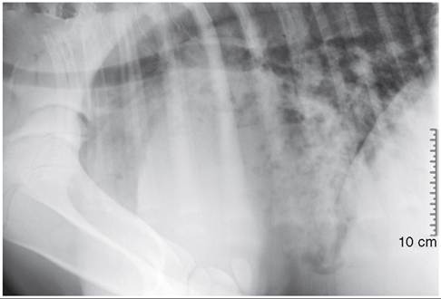
Radiographic assessment of the thorax of large animals remains preferable to ultrasonographic examination for detection of diffuse parenchymal diseases, such as interstitial pneumonia, pulmonary edema, equine multinodular pulmonary fibrosis, fungal pneumonia, acute respiratory distress syndrome (ARDS), chronic disorders, and deep parenchymal or mediastinal abscesses. Radiographic changes can be nonspecific for a particular disease or minimal to nonexistent in diseases such as EIPH or mild IAD. The thorax in adult horses and cattle is filmed as standing lateral, generally requiring a series of three to four separate but overlapping images to capture the entire lung field; thus the benefit of the ventrodorsal view, in which the two lungs may be compared, is lost. Bilateral standing radiographs may aid in localizing lesions to one hemothorax or the other. In neonates and small ruminants the entire lung field can be imaged in one to two views, and as these animals can be more readily handled and restrained, multiple recumbent views are possible. If significant accumulation of pleural fluid is suspected, ultrasonographic examination should be performed first and radiographs obtained following drainage of excess fluid, as fluid may obscure potentially important parenchymal disease.
Four types of radiographic patterns are described for the thorax: alveolar (airspace), interstitial, bronchial, and vascular. In the alveolar pattern, opaque areas coalesce and fully obliterate vessels and bronchi, and air bronchograms may be prominent. This pattern is commonly found in pulmonary edema, pulmonary hemorrhage, equine multinodular pulmonary fibrosis, ARDS, pneumonia with lung consolidation, and neoplasia. Interstitial patterns are the most common pattern noted in equine thoracic radiographs and are characterized by a blurring of the edges of pulmonary vessels, a diffuse increase in lung density, and variable reticular, linear, and nodular opacities. The reticular pattern is most commonly associated with more diffuse infectious lung diseases, pulmonary edema, interstitial pneumonia, and pulmonary fibrosis, whereas the irregular linear pattern is seen most commonly with resolving bronchopneumonia. Nodular opacities are consistent with abscesses, granulomata, and neoplasms. It is rare to see a pure bronchial pattern in a horse; it is usually seen in association with an interstitial pattern. An exception is paired linear opacities or numerous small circular opacities (donuts) representing thickening of large or medium airways in equine bronchitis or bronchiolitis. The vascular pattern is seen in horses radiographed immediately after exercise or in animals with left-to-right cardiac shunts. Finally, extraparenchymal problems such as pleural effusions or free gas may be seen on thoracic radiographs of large animals. Thoracic radiology is far less sensitive than ultrasonography for evaluation of potential rib fractures.
COMPUTED TOMOGRAPHY AND MAGNETIC RESONANCE IMAGING. Advanced radiographic modalities such as computed tomography (CT) and magnetic resonance imaging (MRI) have become increasingly available at academic and private referral practices and can aid in the diagnosis and characterization of certain upper airway disorders in large animal species. Advantages to these systems are that they provide high-resolution three-dimensional imaging and minimize superimposition of overlying structures that may complicate interpretation of skull radiographs. An additional advantage of CT is the high speed of image acquisition. Standing CT units allow for imaging of the head and proximal cervical region in sedated adult horses, thus eliminating the need for general anesthesia. CT provides excellent bone and soft-tissue contrast and is often used in the diagnosis of sinonasal disorders such as dental-related sinus disease, neoplasia, and ethmoid hematomas.17 MRI provides superior soft-tissue detail, and its use is also described for the diagnosis of laryngeal and sinonasal disorders in adult horses.18,19 MRI is currently more limited in its application toward routine use in large animal species because of the availability of equipment, time required for acquisition of images, and potential costs associated with the procedure.
CT imaging has also become a more desirable tool for the diagnosis of pulmonary and extrapulmonary thoracic disease in hospitalized veterinary species, particularly as the considerable reduction in image acquisition time often eliminates the need for general anesthesia. CT thoracic imaging in large animals is limited to small ponies, foals, calves, and smaller species such as camelids, where it has been described in healthy and sick animals with pulmonary and extrapulmonary disease.16,20-22 Imaging of the thorax is not possible in larger animals due to restrictions in currently available CT gantry diameters and limitations associated with exposure settings needed to penetrate a larger torso.
Ultrasonography
Thoracic ultrasonography is useful for diagnostic, therapeutic, and prognostic evaluation of the extraparenchymal thorax, the pleural space, and the peripheral (superficial) parenchyma of the lung and should be considered for complete evaluation of any large animal with pulmonary disease. Unlike thoracic radiography in adult large animals, thoracic ultrasonography is an imaging technique readily available to most practitioners. In many instances it is superior to thoracic radiography; examples include evaluation of pleural effusions, assessment of thoracic trauma and rib fractures, evaluation of neoplasms or granulomata, detection of mediastinal masses or abscesses, and guidance of transthoracic lung biopsy.23-25 Field ultrasonography has also become an important tool in monitoring dairy calves for bovine respiratory disease.26
Ultrasonography is generally performed with the patient standing, although in neonates lateral recumbency may be preferred or even necessary.
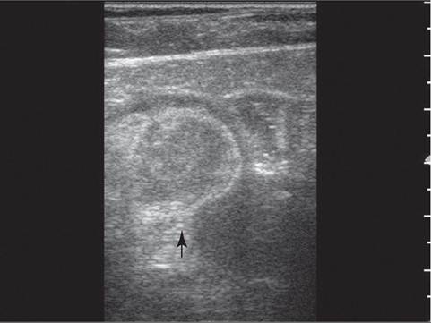
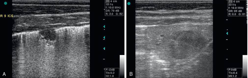
Although ultrasound waves will not penetrate the aerated portion of the lung, limiting the examination to extraparen- chymal surfaces in normal horses, ultrasonography is superior to thoracic radiography for the evaluation of these areas of the chest. Small amounts of pleural fluid that would be missed on auscultation, percussion, or thoracic radiographs can be detected, and the amount and character of pleural effusion in each hemithorax can be evaluated separately.23 Clear fluid is anechoic, but inflammatory cells, gas, and fibrin are echogenic, causing opacities that can be seen floating in pleural fluid and altering the general echogenicity of the fluid. Because of this, ultrasound is the method of choice for diagnosis and monitoring of pleural space disease. Ultrasonography should be used to guide catheter placement for thoracocentesis. The pleural surfaces are imaged well by ultrasound with thickened or roughened areas easily detected. Lack of normal independent movement of the visceral and parietal pleural surfaces during the respiratory cycle, suggestive of adhesion formation, can be readily monitored.23,24
Consolidated lung is a better acoustic medium than aerated parenchyma and can be well visualized. Pleuropneumonia with consolidation or atelectasis caused by compression of the ventral lung by pleural effusion can be evident. Pulmonary abscesses or masses extending to the lung surface can be imaged, and ultrasound can be used for guidance during transthoracic biopsy.23,24 Thoracic radiography remains superior to ultrasound in the diagnosis of pulmonary parenchymal disease and pneumothorax; the two techniques can be combined to optimize diagnostic capacity.
Nuclear Medicine Imaging
Nuclear scintigraphy is a specialized technique available at a few specialty referral practices. Gamma-emitting radioisotopes, most commonly technetium-99m, can be used with an external detector (gamma camera) to assess regional pulmonary ventilation and perfusion in the horse. For perfusion scans, the radioisotope is bound to albumin aggregates and injected into a peripheral vein. The aggregates become trapped in the pulmonary arterial vasculature, and the resulting image illustrates the perfusion distribution of the pulmonary arterial system. The ventilation scan is generated when the horse inhales aerosolized radioisotope particles through a closed circuit system.27 The particles are small enough in diameter to be deposited in the alveoli and small conducting airways, with the gamma camera recording the sites of deposition. Together, the ventilation and perfusion scans allow for evaluation of the ventilation/perfusion (V/Q) ratio, important in evaluation of certain respiratory problems such as chronic EIPH (high V/Q areas), pulmonary thromboembolism (high V/Q areas), and RAO (low V/Q areas). Additional uses are in the evaluation of deposition in the lung of aerosolized radiolabeled medications such as albuterol28 and in the evaluation of mucociliary clearance or tracheal mucus transport where the time required for a bolus of radioisotope to cover a given tracheal distance is evaluated.29
Arterial Blood Gas Analysis
Arterial blood gas measurement is the most sensitive indicator of respiratory function readily available to the clinician. The most easily accessed arteries for sampling are the metatarsal, temporal, facial, and brachial arteries (Fig. 31.2). In cattle the coccygeal artery on the ventral aspect of the tailhead is easily accessible. Heparin is the only acceptable anticoagulant for blood gas samples, and all gas bubbles must be removed and the syringe capped to prevent equilibration of the sample with room air. A short (1-inch), small-gauge (25-gauge) needle and a 1- to 3-mL syringe should be used. Preheparinized syringes with needles can be purchased, or regular syringes and needles may be heparinized by aspirating a small volume of heparin into the syringe via the needle and then forcefully expelling the air and heparin from the syringe three times. This minimizes the effect heparin might have on any reported values from
![]()
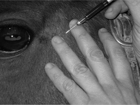
FIG. 31.2 Arterial blood sample drawn from temporal artery for arterial blood gas analysis. (Courtesy Dr. Eric Birks, University of Pennsylvania, New Bolton Center, Kennett Square, Penn.)
■ TABLE 31.1
Normal Arterial pH and PCO2 Values for Various Species (Nonneonate)
| Species | Blood pH | PCO2 (mm Hg) |
| Bovine | 7.32-7.45 | 35-53 |
| Ovine | 7.32-7.54 | 37-46 |
| Equine | 7.32-7.44 | 38-46 |
| Caprine | 7.42-7.46 | 33-38 |
the blood gas analyzer. Pulsation of blood from the needle, spontaneous filling of the syringe, and bright color of the blood all confirm a successful arterial puncture. If arterial puncture is questionable, a comparison sample may be drawn from the jugular vein. Once the sample has been drawn, the vessel should be manually compressed for 2 to 5 minutes to prevent hematoma formation. If the sample will not be analyzed within 10 minutes, it should be placed on ice to slow blood cell metabolism. The patient's body temperature at the time of sampling should also be recorded, as results are frequently reported at both 37° C and as temperature-corrected values at the actual rectal temperature of the patient for pH, PO2, and PCO2 because these values are known to be temperature variable. Clinically, either set of values may be used, but comparisons should be made only between samples reported similarly as either temperature corrected or not. The ranges of normal arterial blood gas values for nonneonates of various species are listed in Table 31.1.
Portable blood gas analyzers are relatively inexpensive and easy to use and provide rapid return of results, making blood gas analysis in large animals more practical in the field.30,31 Awareness of potential device-, species-, or age-related differences in reference ranges, as well as awareness of any limitations in accuracy, is critical for proper interpretation of results. Pulse oximetry is also used in some institutions and referral centers. These monitors measure oxygen saturation of hemoglobin, which is useful for diagnosing severe hypoxemia or recognizing desaturation events, but provide no measure of actual arterial oxygen and carbon dioxide partial pressures; as such, these monitors do not replace arterial blood gas evaluation for assessing adequacy of pulmonary gas exchange.
The most common abnormality recognized with arterial blood gas analysis in an animal that is breathing room air is hypoxemia with normocapnia, hypocapnia, or hypercapnia. Hypoxemia is defined as decreased oxygen tension of the arterial blood (decreased PaO2), whereas hypoxia is defined as decreased oxygen concentration at the level of the tissue, with or without hypoxemia. Hypoxia results from hypoxemia, decreased perfusion of the tissue bed in question, or decreased oxygencarrying capacity of the blood due to anemia or hemoglobin alteration. There are five primary means by which hypoxemia develops in any animal: (1) low partial pressure of oxygen in the inspired air, such as seen at high altitude or in an error mixing ventilator gas (altered FiO2); (2) hypoventilation; (3) V/Q mismatch; (4) diffusion limitation; or (5) intrapulmonary or intracardiac right-to-left shunting of blood. Mild to moderate hypoxemia is not an uncommon finding in neonates, but it must be evaluated in terms of the current age of the foal and its position. The difficulty in obtaining the sample must also be considered, as severe struggling can variably affect the arterial blood gas results.
Hypercapnia (increased PaCO2) develops in response to hypoventilation and may also represent respiratory failure if the lung is significantly involved in the underlying pathology, such as with severe pneumonia or ARDS. It is important to try to distinguish between acute and chronic hypercapnia. Acute hypercapnia is usually accompanied by a relatively dramatic decrease in blood pH of 0.008 pH units for each 1-mm Hg increase in PaCO2. This respiratory acidosis can promote circulatory collapse, particularly in the concurrently hypoxemic and/ or hypovolemic patient. The effects of more chronic CO2 retention are less obvious as the time course allows for adaptation. The pH change is less, about 0.003 pH units per 1-mm Hg increase in PaCO2, as it is balanced by enhanced renal absorption of bicarbonate by the proximal renal tubule. Most patients with acute respiratory distress are in the acute stages of respiratory failure, but chronic adaptation will begin to occur within 6 to 12 hours and will be maximal in 3 to 5 days. An increase in bicarbonate will be noted, particularly if the acidosis is primarily respiratory in origin, and pH may be within the normal range.
Alveolar gas exchange is readily estimated by determining the alveolar-arterial (A-a) gradient for oxygen, computed by subtracting the PaO2 measured by the arterial blood gas from the calculated alveolar oxygen partial pressure (PAO2). The PAO2 is effectively estimated using the partial pressure of inspired oxygen (PiO2) as follows32:
![]()
The PiO2 equals the total barometric pressure (760 mm Hg) minus the partial pressure of water vapor (47 mm Hg) multiplied by the fraction of room air that is oxygen (0.21) and thus equals 150 mm Hg for room air at sea level. For patients on supplemental inspired oxygen, the practitioner must remember to recalculate the PiO2 with the new oxygen fraction (FiO2) (or, if at altitude, the PO2 in the inspired gas), which is only possible in patients receiving inspiratory gas through a closed system. The PaCO2 is obtained from the arterial blood gas measurement. The A-a gradient is normally only 4 to 10 mm Hg; an increase beyond this indicates impaired gas exchange within the lungs, most often the result of V/Q mismatching. Only an estimate of the A-a gradient can be calculated in patients receiving intranasal insufflation of oxygen, as neither the FiO2 nor the PO2 is known.
A second useful measurement is the PaO2ZFiO2 ratio, a component of most definitions of ARDS.33,34 The PaO2ZFiO2 ratio equals the PaO2 obtained from the arterial blood gas divided by the FiO2. The normal PaO2ZFiO2 ratio in most adult animals is greater than 300 mm Hg. In nonneonatal veterinary species, a PaO2ZFiO2 ratio of 201 to 300 mm Hg is consistent with veterinary acute lung injury (VetALI), whereas a ratio of 200 mmHg or less suggests veterinary ARDS (VetARDS). Modified definitions of VetALI and VetARDS exist for neonatal foals (EqNALI and EqNARDS) to account for the relative hypoxemia of normal neonatal foals compared with adults during the first week of life.35 These conditions are described in greater detail in a later section of this chapter (Acute Respiratory Distress Syndrome).Respiratory Function Testing
Numerous techniques have been developed to evaluate respiratory function. Techniques already discussed in this section include arterial blood gas evaluation for assessing efficiency of gas exchange by the lung and videoendoscopy for the qualitative evaluation of upper airway function. Additional techniques that allow for quantitative measurements of upper airway and pulmonary mechanics that influence normal transport of gas from the periphery to the site of gas exchange are available. In most systems, measurements of pressure, flow, and volume allow for the computation of a variety of ventilatory parameters such as respiratory frequency, minute volume, and tidal volume as well as determination of airway resistance and impedance. Pulmonary function testing is performed in horses
Γ φ φ J OL az 'O
for the diagnosis and characterization of equine asthma36-38 and has been described in healthy adult camelids.39,40 These techniques are primarily limited to use as research tools.
A variety of techniques have been described for the evaluation of upper airway mechanics in exercising horses. In addition to acquiring measurements of ventilatory parameters, these systems may also allow for the construction of pressure volume curves to assess the work of breathing or tidal breathing flowvolume loops to detect and characterize airway obstruction.41 Assessment of airway muscle activity using electromyography and respiratory sound analysis have also been described in exercising horses.42,43 One of the major limitations associated with evaluating airway function in exercising horses is the equipment required for these tests. Face masks equipped with a pneumotachograph or pneumotachometer are required to obtain measurements of airflow, whereas nasotracheal or percutaneous tracheal catheters coupled to pressure transducers are used for measurements of airway pressures. These systems may be cumbersome for the exercising horse, may impede achievement of maximal speeds, or may introduce error into results by altering airway mechanics. Recently an ergospirometer has been described that holds promise for acquiring measurements of oxygen consumption (VO2) and ventilatory parameters in exercising horses under field testing conditions.44
Collection and Evaluation of Respiratory Secretions
TRACHEAL ASPIRATES AND BRONCHOALVEOLAR LAVAGE. Various spaces in the respiratory system can be aspirated or lavaged for diagnostic or therapeutic purposes. The most commonly performed procedure is the percutaneous tracheobronchial aspirate. By aspirating from the airways caudal to the larynx, a sample without pharyngeal contamination can be obtained. As discussed previously, endoscopically guided tracheobronchial aspirates can be obtained, and these compare favorably with traditional percutaneous tracheobronchial aspirates provided that a protected aspiration catheter is used.8
In both the horse and the ruminant, the procedure is performed with the animal standing. Sedation or restraint may be needed. A small area over the trachea in the middle third of the neck is clipped and routinely sterilely prepared. The skin is anesthetized using a local block of 2% lidocaine (2 to 3 mL subcutaneously), and a small stab incision is made. A sterile trocar or needle (10-gauge) is introduced on the midline between muscle bundles, with the beveled edge facing ventrally (pointed edge facing dorsally) to decrease the opportunity for inadvertent cutting of the tubing when it is introduced or manipulated, and the ventral tracheal wall is punctured between two cartilaginous rings. The distal end of the trocar or needle is then advanced distally in the trachea, taking care not to lacerate the dorsal tracheal mucosa. Sterile polyethylene tubing or catheter is introduced through the trocar or needle for about 30 cm. A needle or sharp trocar should be withdrawn to prevent severing the tubing or catheter, but a trochar with rounded edges may be left in place. Approximately 20 to 30 mL of nonbacteriostatic sterile isotonic saline solution is introduced quickly. Intermittent aspiration is performed as the tubing is gradually withdrawn. The tubing can be advanced again if a guarding cannula has been left in place to prevent introduction of skin contamination. Additional saline solution aliquots can also be introduced. Once an adequate sample has been obtained, the tubing is completely withdrawn, followed by the withdrawal of the needle or trocar. Commercially available tracheobronchial aspiration kits are also available for use with horses and foals. Injectable antimicrobial solution or suspension can be infiltrated at the skin incision site if a septic sample is suspected, and in horses and small ruminants a sterile dressing can be applied for 24 hours if desired.
Possible complications include subcutaneous emphysema (usually peritracheal but may extend into the mediastinum), local cellulitis, or cutting of the catheter at the needle and loss into the airway. The latter is usually resolved because the catheter is rapidly coughed up, but if necessary, the severed catheter can be retrieved endoscopically. This complication can be readily avoided by using commercially available kits that use a trochar instead of a needle (MILA [MILA International, Elanger, Ky.]), eliminating the needle point that can sever the catheter. The sample should be cultured for aerobic bacteria, and anaerobic cultures should be made if these organisms are suspected based on evidence of pleural effusion, lung consolidation, abscessation, fetid breath, or history of aspiration. For patients with prior antimicrobial therapy, it is advised to discontinue antibiotics for 72 to 96 hours before culture when possible.
A direct smear and Gram stain can be used as an initial guide for antimicrobial therapy pending culture results. Cytologic evaluation can be extremely valuable in differentiating among infectious, allergic, parasitic, and neoplastic processes. If fluid evaluation cannot be performed shortly after collection, it should be stored at 4° C for no more than 24 hours to avoid neutrophil degeneration or bacterial overgrowth. Transtracheal aspirates from clinically normal horses contain columnar ciliated epithelial cells, a few neutrophils, and multiple mononuclear cells. The presence of squamous epithelial cells suggests oropharyngeal contamination, which may occur if the animal coughs during sample collection. Increased percentages of neutrophils and the presence of mast cells, eosinophils, giant cells, and hemosiderophages have been found in aspirates from normally performing Thoroughbred racehorses, indicating some airway inflammation in “normal” equine atheletes.10 Mucus, large spores, and fungal hyphae may be found in the absence of airway disease and must not be overinterpreted. In cases of pneumonia, neutrophils may constitute 40% to 90% of the cellular sample. Bacterial pneumonia causes a more degenerate appearance of neutrophils, and intracellular bacteria may be found. Equine lungworm is characterized by the presence of large numbers of eosinophils and occasionally larvae. In ruminants, the most important information gathered in patients with bronchopneumonia is usually the result of culture and antimicrobial sensitivity testing.
Bronchoalveolar lavage (BAL) involves obtaining a sample from the terminal airways and alveolar region and is described in horses, cattle, and camelids.40,45,46 BAL is performed using a long endoscope (200 to 250 cm) or BAL tube (e.g., doublelumen or cuffed Bivona [Smiths Medical, Norwell, Mass.] or MILA [MILA International, Elanger, Ky.]) introduced through the nares. Endoscopic BAL allows for more exact placement of the end of the endoscope, so a clear understanding of the anatomic location of the distal airway lavage is available. It also allows for the characterization of lower airway secretions and mucosal inflammation. Use of the BAL tube is a blind technique, but most frequently the right dorsal lung is sampled.47 BAL is contraindicated in horses that are expected to exercise within 24 hours of the procedure and in any animal demonstrating signs of respiratory distress, marked tachypnea, paroxysmal cough, or significant hypoxemia.
Proper sedation and restraint is critical for performing BAL. In horses, the addition of butorphanol may reduce cough that is typically associated with this procedure. Instillation of 0.2% to 0.3% lidocaine, particularly when passing the tube into the trachea or past the level of the carina, may also decrease cough. Passage of the tube into the trachea can be facilitated by stretching the head horizontally. The endoscope or tube should be passed until it is wedged in the most distal bronchus. If using a cuffed BAL tube, the cuff should be inflated to seal the airway once wedged. In adult horses, it is recommended to infuse a total of 250 to 500 mL of warm (37° C), sterile physiologic saline in aliquots of 60 to 250 mL. Smaller lavage volumes may be necessary in foals or smaller species. Fluid may be instilled via prefilled syringes or via a solution administration set using a pressure bag with fluid bag or pressure bulb with fluid bottle. The BAL fluid sample is collected via gentle continuous aspiration with syringes or a suction pump using low suction pressure. Cytologic interpretation does not appear to be influenced by method of fluid collection, but syringe aspiration may minimize the potential for barotrauma in certain horses.48 Fluid aliquots recovered should be pooled for analysis. The presence of surfactant (foam) and turbidity on gross inspection is suggestive of a properly obtained sample.
BAL has the advantage of sampling the distal airways, but only a limited area of the lung is sampled instead of the pooled secretions from a tracheobronchial aspirate. Bronchoalveolar lavage is considered superior to tracheobronchial aspirate in evaluation of horses with diffuse chronic lung diseases of the peripheral airways. Molecular analysis of fluid can be performed for identification of certain viral or bacterial species. Cytologic findings of BAL and tracheobronchial aspirates correlate poorly, and the latter should be performed prior to BAL if bacterial pneumonia is suspected. Bronchoalveolar lavage cytology is valuable for evaluation of suspected fungal infections, equine asthma, and EIPH. Similar to tracheobronchial fluid, BAL fluid should be processed within a few hours or stored at 4° C. Normal cell distribution of BAL fluid in healthy horses (based on 250-mL lavage volume) reveals predominantly macrophages (50% to 70%) and lymphocytes (30% to 50%) with lower percentages of neutrophils (in fixative solution.
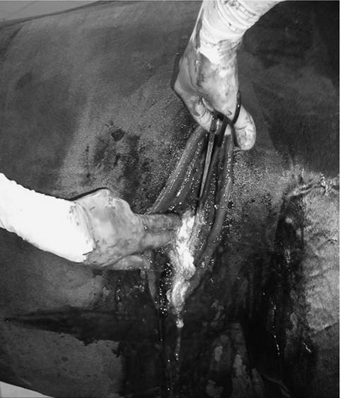
Thoracocentesis
Aspiration from the pleural space is a simple procedure that can be both diagnostic and therapeutic. In the horse with septic or neoplastic effusions, sedation is often unnecessary because the procedure causes only minimal additional discomfort. Following ultrasonographic evaluation of the thorax, a point is chosen where drainage or fluid sampling would seem most appropriate, frequently found in the sixth or seventh intercostal space 10 cm dorsal to the olecranon and above the lateral thoracic vein. The area should be clipped and surgically prepared. Multiple sites may be needed in horses with loculated pockets of fluid in the pleural cavity. The skin and intercostal tissue down to the pleura are anesthetized with lidocaine, and a stab incision is made. A sterile 2- to 3-inch teat cannula or bitch catheter is introduced immediately cranial to the rib border. The cannula should be attached to sterile intravenous (IV) extension tubing and a three-way stopcock. While the cannula is advanced bluntly through the parietal pleura, a sudden loss of the force required to advance is felt. Aspiration should be attempted at this time. The orientation of the cannula can be varied to reach as much fluid as possible. Normally only a few milliliters of straw-colored fluid are obtained. In cases of pleural effusion, as much as 30 L may be removed from each side of the chest (Fig. 31.3). If fluid is excessive, the tubing can be extended over a bucket for gravity drainage, or a vacuum pump with a fluid trap can be attached. When the procedure is complete, a purse-string suture is placed around the stab incision and the cannula is withdrawn while the suture is tightened. In cases where the effusion is large and expected to continue forming for several days, the drainage can be performed by placing a chest tube instead of a teat cannula. If a chest tube is to be left in place, it should be secured with a Chinese finger-trap suture and the end covered by a Heimlich valve to prevent aspiration of air into the thorax through the tube. If the thorax is being drained rapidly, the patient should be watched carefully for signs of distress, as draining of large volumes can alter cardiovascular parameters significantly.
Increasing opacity, presence of fibrin clumps, and malodor of pleural fluid all suggest relative progression from transudate to septic exudate containing inflammatory cells and debris. A
![]()
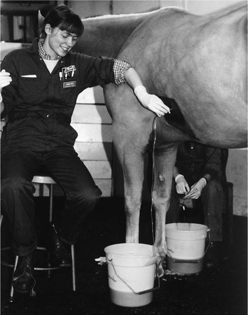
FIG. 31.3 Thoracocentesis and therapeutic drainage in the horse. Pleural effusion can be large and bilateral. Samples should be obtained for culture and cytologic examination at the time the chest is drained. (Courtesy Dr. Corinne Sweeney, University of Pennsylvania, New Bol^ton Center, Kennett Square, Penn.)
putrid odor suggests the presence of anaerobic bacteria. Samples should be cultured for aerobic and anaerobic organisms. A white blood cell (WBC) count of 10,000/mL or less is considered normal; fewer than 60% are normally neutrophils, the remainder being lymphocytes and macrophages. The proportion and total number of neutrophils increase with pleuritis. Erythrocytes are normally not present in the absence of a traumatic tap. The protein concentration is normally less than
3.5 g/dL, and pH should be approximately 7.4. Additional metabolic values that give early indication of sepsis can be obtained on pleural fluid samples collected after filtration through a blood administration set to remove fibrin and debris potentially detrimental to analytic equipment. Pleural fluid pH, PCO2, and concentration of glucose, lactate, and bicarbonate can be directly compared with similar analysis of venous blood from the patient. A septic pleural exudate is acidic, with decreased glucose and bicarbonate but increased lactate and PCO2 compared with venous blood concentrations or tensions, apparently reflecting metabolic activity of phagocytic cells and bacteria and development of an anaerobic environment.49 Of these values, low pleural fluid glucose concentration (drops, facilitating drainage of the secretions by gravity. A Chambers mare catheter can be passed blindly through the ventral meatus into the pharynx. The curved end is directed beneath the flap of the medial lamina of the pouch ipsilateral to the nostril used for passage. Successful passage is indicated by lack of resistance while the catheter is inserted deeper than if it were in the pharynx. The position of the catheter tip in the pharynx can be observed through an endoscope placed up the opposite nasal passage. Once the catheter is within the pouch, it can be used to obtain a sample, to drain excessive secretions, or to act as a conduit for flushing. A self-retaining uterine catheter can be left in place for repeated flushing, but the Chambers catheter can be passed repeatedly with no complications.
Airway and Lung Biopsy
Biopsy of the airways or lung is most often performed in the horse and is indicated to obtain a histologic diagnosis or prognostic information primarily in cases of diffuse lung or airway disease. Endobronchial biopsy is performed for the collection of airway samples, whereas parenchymal samples are obtained by means of percutaneous or thoracoscopically guided collection techniques. For endobronchial biopsy, multiple samples can be collected along the upper and lower airways. Histologic analysis of endobrochial samples collected from peripheral airways may have clinical utility for diagnosis of RAO in horses.51 Thoracoscopically guided pulmonary wedge resection is also an option for collection of a larger parenchymal tissue sample and enables direct visualization of the lung prior to biopsy. The procedure is relatively safe but is more invasive and requires specialized equipment and expertise.14 For percutaneous biopsy, use of a spring-loaded biopsy instrument with a large-gauge (14-gauge), 15-cm or longer biopsy needle and a sample “slot” length of 22 mm is recommended. Discomfort is minimal, and sedation may or may not be needed. The site for biopsy should be determined following ultrasonographic evaluation and should be away from common locations of major pulmonary vessels. Caudal and dorsal locations are generally chosen. The site should be surgically prepared and infiltrated with local anesthetic down to the pleura. The biopsy needle is inserted through a stab incision just cranial to the rib and directed medially through the intercostal muscles and parietal pleura. The needle should be advanced the distance indicated by ultrasonographic measurement, then sharply advanced less than 1 cm to enter the lung parenchyma and the spring-loaded instrument “fired” to obtain the sample; the biopsy instrument is then withdrawn. After sufficient biopsy specimens have been obtained, a single skin suture can be placed at the incision site, but no additional aftercare is needed. A specimen should be placed directly in 10% formalin or glutaraldehyde for fixation with additional samples submitted for bacterial and fungal culture, and potentially for more advanced molecular diagnostic techniques. Complications of lung biopsy have been reported to range from transient epistaxis or hemoptysis, which is to be expected, to more severe pleural and parenchymal hemorrhage. Lung biopsy is not indicated for pleuropneumonia cases but is generally required to differentiate between equine multinodular pulmonary fibrosis, discrete fungal lesions, and potential neoplasia, when accurate diagnosis is required for therapeutic and prognostic purposes.
Techniques for Identification of Respiratory Pathogens
Infectious respiratory disease can have significant individual and herd health implications. Timely and definitive identification of the causative agents is necessary to ensure that the appropriate therapeutic, prophylactic, and biosecurity protocols are instituted. Although infectious respiratory disease may represent a diagnostic challenge for the equine practitioner, technological innovations have improved the speed and sensitivity with which infectious organisms can be identified.52 Culture, immunologic tests, and molecular techniques represent the most commonly used tools for pathogen identification. These techniques should not substitute for careful clinical evaluation, and an accurate diagnosis may necessitate the use of more than one diagnostic test. Regardless of the technique chosen, the practitioner should adhere to appropriate guidelines for sample collection and handling, as failure to do so can negatively affect the diagnostic accuracy of a given test.
Conventional culture methods represent indispensable diagnostic techniques when infectious respiratory disease (bacterial, viral, fungal) is suspected. Although these methods are sensitive, they are time consuming, often delaying pathogen identification days to even months, as in the case of certain viral organisms. Variable recovery of organisms from respiratory secretions and contamination may also negatively affect diagnostic accuracy. Matrix-assisted laser desorption/ionization- time-of-flight mass spectrometry (MALDI-TOFMS) is a novel technology that uses pathogen-specific mass spectra ribosomal protein profiles for the identification of fungal and microbial pathogens.53 Extensive databases of organism-specific MALDI- TOFMS profiles have been generated, and when compared to conventional culture, MALDI-TOFMS can reduce the time required for pathogen identification from days to hours. This has important clinical implications regarding more timely institution of appropriate management.
IMMUNOLOGIC TECHNIQUES. Immunoassays rely on interactions among bacterial, viral, and fungal antigens and radioisotope-, enzyme-, or fluorescent-labeled antibodies. The use of polyclonal antibodies tends to increase the sensitivity of the assay, as the preparation may contain antibodies to multiple epitopes on the target antigen, but at the same time it may also decrease assay specificity due to their heterogeneous nature. Test specificity can be improved by the use of monoclonal antibodies, as these antibodies interact with only a single well-defined epitope or very similar epitopes. Immunofluorescence, although labor intensive and time consuming, is considered highly sensitive and specific, particularly for virus- infected cells. The enzyme-linked immunosorbent assay (ELISA) is available for the diagnosis of a wide range of infectious respiratory diseases. For certain pathogens the ELISA has the advantage of rapid turnaround of results and minimal labor; however, these tests may be subject to false-negative and false-positive results. Virus neutralization (VN) assays are highly accurate and sensitive. Unfortunately, VN is more expensive, labor intensive, and time consuming compared to other diagnostics.
Immunohistochemistry is a standard diagnostic tool for the identification of viral, bacterial, and protozoal pathogens in tissue sections. This technique depends on polyclonal or monoclonal antibodies binding to a target antigen and the demonstration of this interaction by colored histochemical reactions visible by light microscopy or by emittance of fluorescence. Immunohistochemistry is highly versatile, and assays have been developed to detect a variety of equine respiratory pathogens in tissue sections.
MOLECULAR TECHNIQUES. Molecular-based technology, in particular polymerase chain reaction (PCR) techniques, have greatly advanced the diagnosis and management of viral, fungal, and bacterial respiratory disease. Advantages to these techniques include the speed with which results can be obtained, the superior sensitivity and specificity of these assays, and the ability to obtain quantitative results. Nucleic acid of both live and dead pathogens can be detected at very low concentrations and in a wide variety of specimens, and PCR is often useful to identify organisms that are difficult to grow in culture. The ability to perform parallel (“panel”) testing for multiple respiratory pathogens simultaneously from a single sample has greatly increased diagnostic efficiency, particularly when co-infection of multiple pathogens exists. Other advances that have expanded the clinical utility of PCR techniques include identification of virulence-associated genes, discrimination among different viral strains as well as between viable and nonviable organisms, determination of infectious risk, and the ability to simultaneously test for multiple genes from the same organism.
Polymerase chain reaction involves amplification of a target region of DNA. Advances in PCR technology such as reverse transcription PCR (RT-PCR) and real-time, or quantitative, PCR (qPCR) have greatly expanded the applications of standard PCR. RT-PCR enables identification of viruses whose genome is composed of RNA (e.g., influenza, coronavirus) by reverse transcribing the viral RNA into complementary DNA (cDNA) for amplification. Commercially available kits have increased the diagnostic utility of this technique. Real-time PCR (qPCR) combines PCR amplification with simultaneous detection of the amplified products, allowing for quantification of PCR products in a closed tube system. Real-time PCR may also be combined with RT-PCR (qRT-PCR) for the quantification of messenger RNA (mRNA). Compared with traditional PCR, qPCR reduces the risk of contamination and increases the sensitivity and speed of detection.
Adhering to appropriate sample collection and handling protocols is essential for diagnostic accuracy of PCR. For example, sterile synthetic nasal or nasopharyngeal swabs (rayon or dacron tipped) are recommended for viral identification, whereas nasopharyngeal or guttural pouch lavage is recommended for identification of Streptococcus equi subsp. zooepi- demicus. Regardless of the source, samples should not be frozen but refrigerated at 4° C for no more than 2 to 3 days before processing. False-positive results are often due to contamination, and false-negative results may occur from enzyme inhibitors that suppress DNA amplification. Molecular diagnostic techniques are covered in greater detail in Chapter 29.
Novel techniques that have the potential to further our understanding of the pathogenesis as well as the immunologic and genetic bases of respiratory disease include flow cytometry, genome-wide association study (GWAS), and respiratory microbiome analysis. Although these techniques currently are primarily used as research tools, they may have broader clinical applications in the future by contributing to improvements in the prevention, diagnosis, and management of respiratory disease. Flow cytometry is a laser-based technology that detects and quantifies light scatter and fluorescence emitted by fluorescent-labeled cells. Combinations of scattered and fluorescent light are identified and analyzed to derive information about the physical and chemical properties of a large number of cells at one time. It can be used to immunophenotype cells isolated from respiratory secretions and may thus improve characterization of the immunologic response associated with select respiratory diseases.
The goal of a GWAS is to identify genomic features that may influence the risk of disease or the pathogenesis of a disease in animals or humans. The entire genome is evaluated in individuals with and without a particular disease to identify specific genetic variants found more frequently in individuals with disease. It is important to recognize that a GWAS does not determine causation, but only association. It may, however, facilitate identification of candidate genes that can be further investigated. In large animal species GWAS is being used to improve the understanding of genetic influences on susceptibility to severe RAO in warmblood horses,54 equine viral arteritis in stallions,55 and bovine respiratory disease in cattle.56
The role that the respiratory tract's resident microbial communities may have on respiratory health has received considerable attention through the field of respiratory microbiome analysis. In particular, the respiratory microbiome is thought to contribute to the prevention of colonization by pathogenic organisms, the mitigation of pathogenicity when infection is established, and the structural and immunologic maturation of the respiratory tract.57 Important areas of ongoing respiratory microbiome research in large animals include characterization of different microbial communities throughout the respiratory tract, identification of factors influencing the development of the microbiota in maturing animals, and understanding how certain events (e.g., transport, physiologic stress, antimicrobial or glucocorticoid administration) may alter the microbiome and potentially influence susceptibility to or pathogenicity of respiratory disease.58-61
Pulmonary Function Testing
Daniela Bedenice
Pulmonary function testing (PFT) has emerged as an essential tool in equine referral practice and is largely aimed at describing the severity, anatomic pattern, stability (reactivity), and reversibility of bronchoconstriction caused by noninfec- tious airway obstruction and inflammation, such as is seen in RAO and IAD.1 Clinical indications for noninvasive PFT include assessment of horses with intermittent cough, exercise intolerance, abnormal breathing pattern, and excess mucus, as well as early detection of subclinical disease, evaluation of treatment response, intensive care monitoring, and prediction of outcome. PFT is generally divided into the assessment of respiratory mechanics (mechanical properties of the respiratory system) and gas exchange. Analysis of gas exchange investigates ventilation-perfusion matching, shunt, diffusion capacity, and dead space-to-tidal volume ratio (Vd∕Vt). Lung mechanics, on the other hand, determine the static and dynamic properties of the lung, including resistance, compliance, functional residual capacity (FRC), and ventilatory parameters.1
Mechanics of Breathing
The mechanical function of the lung is essentially defined by static and dynamic properties. Tests that are performed with the respiratory system at equilibrium and zero flow are referred to as static tests. Examples include measurement of lung volume subdivisions (e.g., FRC) and compliance of the lung and chest wall.1
FRC is a measure of the amount of gas that remains in the lungs at end-expiration (end-expiratory volume). FRC is lower in patients with increased “lung stiffness” (reduced elastic recoil of the lung) as well as airway inflammation, whereas FRC increases in patients with expiratory airway obstruction and gas trapping.2 This test can be easily performed in awake clinical patients via a helium dilution technique.3 In short, the patient is connected to a reservoir bag at end-expiration to rebreathe a standard, commercial breathable gas mixture of 10% helium (He), 20% oxygen, and the remainder nitrogen for 90 seconds. The dilution of He (a nonexchangeable gas) gives a measure of end-expiratory lung volume.3
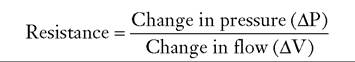
Measurements of static compliance or “elastic recoil of the lung” require breath holds and have applications only to the anesthetized patient. So-called pressure-volume curves are generated in the relaxed patient during lung deflation from total lung capacity (TLC), using an esophageal balloon technique. Compliance is defined as the lung volume change per unit of 2
pressure change2:
![]()
Reduced lung compliance (i.e., increased lung stiffness) may be associated with increased fibrous tissue (pulmonary fibrosis), atelectasis (e.g., underventilated lung), or an increased pulmonary venous pressure, in which the lung becomes engorged with blood. Emphysema and normal aging of the lung, which leads to alteration in elastic tissue, are causes of increased lung compliance.2
In contrast to static tests, tests that are performed with the respiratory system in motion (e.g., during quiet breathing) are referred to as dynamic. An example of a dynamic measurement is resistance, a measurement that requires flow. Resistance arises from friction of air molecules against airway walls1:
![]()
The measurement of pulmonary resistance (RL) using a flow meter attached to a face mask and an esophageal balloon catheter to measure transpulmonary pressure changes is conventional in the horse but rarely used in the clinical setting.1 This technique allows computation of both Rl and dynamic compliance (Cdyn) at spontaneous breathing frequencies. Maximal transpulmonary pressure change (ΔPPLmax) and Rl both increase in cases of obstructive airway disease, whereas Cdyn decreases. However, this classic technique is fairly insensitive in detecting subclinical disease4,5 and has greater utility in the diagnosis of IAD if coupled with a challenge test (i.e., histamine bronchoprovocation [see below]).6,7
Forced Oscillation Techniques
In contrast to the conventional methods, noninvasive measurements of total respiratory system resistance via forced oscillation techniques (FOTs) are used in the diagnosis of IAD in horses. In short, oscillometry is the study of lung mechanical function via the application of external forces to the respiratory system.8 Either a loudspeaker (e.g., impulse oscillometry system [IOS]) or air pressure (e.g., monofrequency forced oscillation) is used to superimpose pulses of flow (4 to 5 L/s peak) through a face mask on the horse's respiratory system during spontaneous breathing. The generated reciprocal pressure waves are subsequently recorded at the airway opening (i.e., face mask). The magnitude and phase relationship between the input of flow and output of pressure are then used to perform the calculations of impedance (total opposition to airflow) and its components, resistance and reactance, at a variety of oscillatory frequencies (generally 1 to 7 Hz).1 In most horses with IAD (equine asthma) there is a frequency dependence of resistance, with higher values for resistance recorded at the lower oscillatory frequencies (1 to 2 Hz), indicative of bronchoconstriction.9-11 Horses with moderate to severe lower airway obstruction commonly show high initial baseline values for respiratory resistance at 1 Hz (e.g., >1.0 cm H2O∕L∕s).1 Higher frequencies (≥2 Hz) provide information concerning central airway resistance (Raw). Baseline respiratory resistance measurements using FOTs are often combined with bronchoprovocation tests for the early diagnosis of IAD. In addition, the use of an IOS has been advocated for separate analysis of inspiratory and expiratory impedance parameters, to allow the evaluation of both phases of the respiratory cycle in horses.12
Histamine Bronchoprovocation
Bronchoprovocation is a challenge test that assesses the response of the respiratory system to a bronchoconstrictor agonist (e.g., inhaled histamine).1 The provocatory concentration necessary to cause a 100% increase in baseline respiratory system resistance is commonly termed PC100RRS. Horses with a low PC100rrs (e.g., is broad and reflects the severity of the disease process. Early identification of affected animals and immediate initiation of appropriate therapy are essential to prevent mortality and functional impairment of the respiratory system.
Infectious Agents Involved
Adult horses most commonly acquire bacterial pneumonia by aspiration of microorganisms that normally inhabit their nasopharynx or oral cavity.1,2 β-hemolytic streptococci, particularly Streptococcus equi subsp. Zooepidemicus (S. Zooepidemicus) is by far the most common bacterial pathogen isolated from adult horses with bronchopneumonia.3,4 Nonenteric gramnegative bacteria such as Pasteurella spp. and Actinobacillus spp. are also frequently isolated, either alone or in combination with S. zooepidemicus. Enteric gram-negative bacteria such as Klebsiella spp., Escherichia coli, Enterobacter spp., and Salmonella enterica may also be isolated. Other aerobic gram-positives such as Staphylococcus spp. and Rhodococcus equi or gram-negatives such as Pseudomonas spp. and Bordetella bronchiseptica are occasionally isolated. Pseudomonas spp. are rarely a primary cause of pneumonia in horses, and their presence often reflects contamination of equipment used for taking airway samples (such as endoscopes). Streptococcus pneumoniae, a common pathogen of humans, has been positively correlated with lower airway inflammation in young Thoroughbred racehorses in the United Kingdom.5 The microorganism can also induce pneumonia in ponies following heavy intrabronchial challenge.6 Its isolation from pneumonic horses in the United States appears to be rare.
Anaerobic bacteria are isolated from approximately one third of adult horses with severe bronchopneumonia, pleuropneumonia, or pulmonary abscessation. The most common anaerobes isolated are Bacteroides spp. (particularly Bacteroides fragilis), Clostridium spp., and Peptostreptococcus spp.; Fusobacterium spp. and Eubacterium spp. may also be isolated.3,7 Isolation of anaerobes from horses with pneumonia or pleuropneumonia has been associated with a less favorable prognosis in some studies. In one study, the survival rate for 221 pneumonic horses with strictly aerobic isolates from tracheobronchial aspirates was 81.4% compared with 38.3% for the 81 horses in which anaerobes were cultured.3 Mixed bacterial infections are very common and may represent synergy between aerobic or facultative aerobic and anaerobic bacteria.
The importance of Mycoplasma spp. in the development of equine bronchopneumonia and pleuropneumonia is controversial. Several Mycoplasma species have been isolated from the respiratory tract of both diseased and healthy horses, with Mycoplasma felis and Mycoplasma equirhinis being the most common isolates. In one study, isolation of M. equirhinis was positively correlated with lower airway inflammation in a group of young Thoroughbred racehorses in the United Kingdom,8 whereas isolation of Mycoplasma spp. was not significantly associated with disease in a similar population in Australia.9 An outbreak of lower respiratory tract disease caused by M. felis infection has been described.10 M. felis has also been isolated from horses with pleuropneumonia, and experimental infection with M. felis has resulted in pleuropneumonia.11,12
■ Epidemiology Bacterial bronchopneumonia may affect horses of any age and breed. In one retrospective study of 327 horses with pneumonia or pleuropneumonia, there was no sex predilection but 82% of the horses were younger than 5 years of age.3 In a retrospective case-control study of risk factors for development of pleuropneumonia, Thoroughbreds were at greater risk and Standardbreds were at lower risk of developing the disease.13 In the same study, the most significant risk factor for development of pleuropneumonia was long-distance transport within the week prior to the onset of clinical signs.13 In another study, 24.4% of 90 horses with pleuropneumonia had recently been transported over long distances, and 12.2% had recently undergone general anesthesia.14 Five of the postsurgical cases had undergone upper airway surgery.14 Whether these horses had preexisting lung disease or whether they developed aspiration pneumonia as a result of surgery and general anesthesia could not be ascertained.
Other factors significantly associated with increased risk of developing pleuropneumonia include recent viral respiratory tract infection or exposure to horses with viral infections and racing within 48 hours prior to developing clinical signs.13 One study in the United Kingdom identified a higher incidence of pneumonia or pleuropneumonia in show jumpers, presumably reflecting the greater distance over which these horses are transported compared to racehorses in that country.15
■ Pathophysiology Colonization of the lungs by opportunistic bacteria occurs when the pulmonary defense mechanisms are compromised or are overwhelmed by massive numbers of bacteria. Several factors can contribute to causing increased numbers of bacteria in the lower airways. Dysphagia or esophageal obstruction will lead to aspiration of large numbers of pharyngeal bacteria, and these disease processes often result in pneumonia. However, the vast majority of horses with bacterial pneumonia or pleuropneumonia do not have a history of dysphagia or esophageal obstruction. Other factors that have been shown to significantly increase bacterial contamination of the lower respiratory tract include confinement with the head elevated, transportation, and high-intensity exercise.16-18 In one study, confinement of horses with the head elevated resulted in a significant increase in bacterial numbers as well as neutrophilic inflammation in the lower respiratory tract as early as 6 hours after initiating confinement.17 Actinobacillus spp., Pasteurella spp., and β-hemolytic streptococci were the predominant bacterial isolates. Lowering the head for 30 minutes every 6 hours to facilitate postural drainage during a 24-hour confinement did not prevent multiplication of bac- teria.17 Clearance of accumulated secretions and bacteria occurred within 8 to 12 hours after release from confinement.17 In similar confinement experiments, pretreatment with penicillin considerably reduced the number of β-hemolytic streptococci but did not reliably reduce total bacterial numbers.19 Cilia of many other species such as dogs and humans can transport mucus effectively against gravity, and posture has no effect on tracheal mucociliary transport in these species.20,21 In contrast, periods of lowered head posture are absolutely essential for normal mucociliary clearance in horses.22
In one study, long-distance transport by road over 12 hours resulted in increased bacterial contamination and neutrophilic inflammation in tracheal aspirate fluid when the horses' heads were restrained in an elevated position.23 This is in contrast to another study in which there was no significant cytologic or bacteriologic changes in BAL fluid from mares whose heads were not restrained in an elevated position during transportation for 12 hours.24 Collectively, these findings suggest that duration of time with a raised head position is more important than the stress of transport alone in development of airway colonization. Finally, the last factor shown to increase bacterial contamination of the large airways of horses is exercise. In one study, a single bout of high-intensity exercise resulted in a tenfold increase in aerobic and hundredfold increase in anaerobic bacterial counts in tracheal aspirate samples compared to preexercise values.16
Pulmonary defense mechanisms can be compromised by numerous factors. These include stress (e.g., transport, intense exercise), viral infections, malnutrition, exposure to dust or noxious gases, immunosuppressive therapy, immunodeficiency disorders, and general anesthesia. Infections with influenza virus and equine herpesvirus 4 (EHV-4) have been shown to significantly decrease mucociliary clearance for up to approximately 30 days post infection in horses.25 In one study, longdistance transport resulted in a significant reduction in phagocytosis by peripheral blood neutrophils for approximately 36 hours after transport.23 The effects of long-distance transport on number, viability, and function of cells in BAL fluid have given conflicting results, with no clear pattern emerging.24,26-28 Similarly, the effects of exercise on the immune system are complex and depend on a multitude of factors, including the intensity or duration of exercise, the specific immune function being analyzed, and the timing of the measurement in relation to exercise. In general, moderate exercise enhances immune function, whereas strenuous exercise tends to be detrimental to immune function.29 High-intensity exercise in horses leads to a significant decrease in blood neutrophil and bronchoalveolar macrophage phagocytic activity, which may lead to decreased bacterial clearance from the lungs.30-32 Exercise also affects the adaptive immune system, as lymphocyte proliferation responses and interferon (IFN)-γ production are decreased following 3335
high-intensity exercise.33-35 Strenuous exercise has also been shown to increase susceptibility to experimental influenza virus infection in ponies compared with rested animals.33
Regardless of the exact mechanism predisposing to bacterial colonization, the inflammatory response induced by bacterial invasion results in infiltration with neutrophils and other inflammatory cells into the airways and pulmonary parenchyma. Inflammatory cells and their mediators cause damage to the airway epithelium and capillary endothelium, leading to flooding of the terminal airways with inflammatory cells, serum cellular debris, and fibrin. This process is generally more severe in the ventral portions of the lung. In early stages the lungs may be simply edematous, reflecting inflammation and early exudation, whereas in chronic, severe cases the airways may be consolidated or abscessed. These lesions interfere with gas exchange, and if severe enough, the resulting ventilation-perfusion mismatch leads to hypoxemia and clinical signs of respiratory disease.
In severely affected animals, inflammation will extend to the pleural space. The first stage of bacterial pleuropneumonia is an exudative stage, which is characterized by rapid outpouring of sterile pleural fluid into the pleural space in response to inflammation of the pleura. The associated pneumonic process is usually contiguous with the visceral pleura and results in increased permeability of the capillaries in the visceral pleura. If appropriate antimicrobial therapy is initiated at this stage, the pleural effusion may not become septic and may resolve. With progression, the bacteria invade the pleural fluid from the contiguous pneumonic process and the second, fibropu- rulent, stage evolves. This stage is characterized by the accumulation of large amounts of pleural fluid with many degenerate neutrophils, bacteria, and cellular debris. Fibrin is deposited in a continuous sheet, covering both the visceral and the parietal pleura adjacent to affected areas. As this stage progresses, there is often loculation and formation of limiting membranes. These loculations prevent dissemination of the infection but make drainage of the pleural space with chest tubes increasingly difficult. The last stage is the organization stage, in which fibroblasts grow into the exudate from both the visceral and the parietal pleura surfaces and produce a membrane called the pleural peel. This inelastic pleural peel encases the lung and renders it virtually functionless. At this stage the exudate is generally thick.
■ Clinical Signs and Physical Examination The spectrum of clinical signs shown by horses with bacterial lung infection is broad and usually reflects the severity of the disease process. Horses with septic IAD and no or minimal involvement of the lung parenchyma may be completely normal at rest. Clinical signs may be limited to exercise intolerance and/or poor performance, or affected horses may cough or have bilateral nasal discharge during or immediately following exercise. Even in early cases of bronchopneumonia, clinical signs may not be obvious. As the disease progresses, clinical signs may include any combination of fever, anorexia, depression, bilateral nasal discharge, cough, weight loss, tachypnea, and respiratory distress. Nasal discharge is usually mucopurulent but may be hemorrhagic in some cases with pulmonary infarction and necrotizing pneumonia.36 Halithosis and a foul-smelling nasal discharge may be present and have been associated with infection by anaerobic bacteria. However, the absence of a putrid odor does not rule out infection with anaerobic bacteria.
Because the parietal pleura is highly innervated and painful when inflamed, horses with acute pleuropneumonia often exhibit pleurodynia (pleural pain). Pleurodynia can often be detected by applying digital pressure to the intercostal space, resulting in grunts, intercostal muscle spasm, or even escape maneuvers by the patient when pain is present. Pleurodynia may also be manifested as pawing, stiff forelimb gait, abducted elbows, and reluctance to move. The condition may easily be mistaken for colic, exertional rhabdomyolysis, or laminitis. As more fluid accumulates in the pleural space and the disease becomes chronic, pain is less evident. A plaque of sternal edema is a common clinical finding in horses with pleuropneumonia. This is not a specific finding, as it can occur with many other disease processes.
Careful auscultation after application of a rebreathing bag (if not precluded by respiratory distress) is extremely valuable in defining the presence and sometimes extent of lung involvement. Horses with a large amount of secretions in the trachea often have an audible tracheal rattle. Most horses with bronchopneumonia will cough when the rebreathing bag is applied, whereas horses without bronchopneumonia will not. Occasional inspiratory or expiratory crackles and/or wheezes may be heard over affected areas, which are more commonly located ventrally. Because consolidated lung parenchyma is a good acoustic medium, mild consolidation sometimes results in only increased bronchial sounds. In contrast, the lung sounds may be diminished in areas of severe consolidation, extensive abscess formation, or pleural effusion.
Auscultation of horses with pleuropneumonia reveals normal lung sounds in the dorsal lung fields, with no sound or considerably decreased lung sounds ventrally. Pleural friction rubs are often not heard because they are present only in the acute stage of the disease. If they are heard, friction rubs are present predominantly at the end of inspiration and the early part of expiration. They disappear as inflammation decreases or as pleural fluids accumulate. Cardiac sounds are often heard over a wider area of the chest than normal, probably as a result of enhanced conduction of sound through the pleural fluid. Thoracic percussion is useful to detect and delineate pleural effusion with resonant sounds dorsal and dull sounds ventral to the horizontal line of effusion.
■ Differential Diagnosis When physical examination and auscultation indicate pulmonary disease, the major clinical task is differentiating infectious from noninfectious causes. Among infectious causes, bacterial pneumonia is most common. Most viral infections are confined to the upper respiratory tract. The clinical presentation of equine multinodular pulmonary fibrosis may resemble that of horses with bacterial bronchopneumonia. Pulmonary aspergillosis most often follows severe gastrointestinal (GI) disease that had resulted in mucosal compromise.37 Severe respiratory disease unresponsive to antimicrobial therapy might suggest fungal pneumonia or equine multinodular pulmonary fibrosis. Parasitic pneumonitis is rare in adult horses but may be seen in horses pastured with donkeys or mules. The two major noninfectious causes of pulmonary disease that may be confused with bacterial pneumonia include heaves (also known as RAO) and IAD. Other uncommon causes of noninfectious pulmonary disease that may result in similar clinical signs are pneumothorax, pulmonary edema, smoke inhalation, neoplasia, and idiopathic interstitial pneumonia. The diagnostic tests described below aid in the differentiation between infectious and noninfectious causes of lower respiratory tract disease.
When pleural effusion is present, several differentials should be considered. Bacterial pleuropneumonia is by far the most common cause of pleural effusion in horses. In one study, 90 of 122 horses (73.8%) with pleural effusion had pleuritis secondary to bacterial pneumonia or lung abscessation.14 Other less common causes of pleural effusion in horses include hemothorax, penetrating chest wounds, esophageal ulceration or rupture, neoplasia, fungal pneumonia, pericarditis, congestive heart failure, diaphragmatic hernia, hypoproteinemia, and chylothorax.
■ Diagnostic Approach Presumptive diagnosis of lower respiratory tract infection is generally based on clinical signs and careful auscultation of the lungs with a rebreathing bag. The need for additional diagnostic procedures is determined by the severity and duration of clinical signs, the number of affected animals, the value of the affected animals, and prior treatment used and response to such therapy. Bacterial pneumonia or pleuropneumonia most commonly affects an individual horse on a given farm. In horses suspected of having bacterial pneumonia, the goal of diagnostic evaluation is to rule out diseases of the upper respiratory tract and to determine the cause and severity of lung involvement.
HEMATOLOGY AND BIOCHEMISTRY. Horses with bacterial bronchopneumonia commonly have an inflammatory hematologic profile. Leukocytosis and absolute neutrophilia with or without a left shift are supportive of bacterial infection. Neutropenia with a toxic left shift may also be evident in the acute stages in severely affected animals. Increased fibrinogen and/or serum amyloid A concentrations, hyperglobulinemia, and mild hypoalbuminemia are common. Anemia of chronic inflammation may develop in chronically affected animals. There does not seem to be a good correlation between the severity of clinical signs and the presence or the magnitude of hematologic changes. When plasma fibrinogen concentrations are increased, their sequential measurement provides a useful means of monitoring response to treatment. It must be remembered that a normal hematologic profile does not rule out bacterial bronchopneumonia.
ENDOSCOPY. Endoscopy may be useful to rule out upper airway infection in situations where physical examination and auscultation of the lungs are not conclusive. Presence of mucopurulent secretions in the trachea and bronchi confirms lower respiratory tract disease. However, the presence of mucopurulent secretions does not necessarily indicate a bacterial infection, as this is also a common finding in horses with RAO and IAD. Bronchoscopy may also be useful in locating the affected lung segment in horses with focal pneumonia or draining pulmonary abscesses. It should be noted that exploration of the trachea and lower airways via bronchoscopy may contaminate subsequent samples taken for culture.
TRACHEOBRONCHIAL ASPIRATE FOR CYTOLOGY AND CULTURE. Tracheobronchial aspirate (TBA) for cytologic examination and bacterial culture is one of the most helpful diagnostic procedures available when bronchopneumonia is suspected. BAL is not as useful as TBA in cases of bronchopneumonia because with BAL, only a small portion of a lung is sampled. In contrast to BAL, TBA samples contain a pooled sample of secretions from all portions of the lung, thus increasing the chances of culturing the pathogen of interest. In one study involving 22 horses diagnosed with pneumonia or pleuropneumonia based on thoracic radiographs or ultrasonography, BAL cytology was abnormal in only 10 horses despite attempts at selectively sampling the affected area.38 In contrast, all horses had evidence of septic inflammation based on tracheobronchial aspirate cytology.38 Whenever possible, antimicrobial therapy should be discontinued at least 24 hours before performing TBA. TBA is preferably obtained by sterile percutaneous transtracheal aspiration to avoid contamination from the upper airway. Alternatively, the sample can be collected with a sterile guarded aspiration catheter passed through the biopsy channel of a flexible endoscope.39,40 Endoscopy has the advantage of allowing selective aspiration of exudate when it is present, which may therefore enhance recovery of bacteria. However, bacterial contamination of nasal or pharyngeal origin more commonly occurs in TBA samples collected via endoscopy. Airway fluid specimens should be submitted for cytology, Gram stain, and aerobic and anaerobic bacterial cultures. The fluid used for anaerobic cultures should be transferred to the laboratory immediately after collection in a manner that prevents or minimizes exposure to air. Anaerobic transport media are commercially available and should be routinely used. Specimens submitted for isolation of anaerobes should not be refrigerated because many anaerobes are intolerant to cold. In horses with pleural effusion, TBA should be obtained even if pleural fluid is available for bacterial culture. In one study, culture of the pleural fluid was negative in 43% of 111 horses with pleuropneumonia, whereas tracheobronchial fluid yielded growth in all cases.3 Only approximately 5% of cases had growth from the pleural fluid but not from TBA.3
Macrophages and columnar ciliated epithelial cells predominate in TBA from healthy horses. The percentage of neutrophils in TBA from apparently healthy horses can be quite variable. In one study, approximately 75% of apparently healthy Thoroughbred racehorses in training had less than 20% well- preserved neutrophils.41 Occasional plant spores and fungal hyphae may be present either free or in large mononuclear cells. Their presence does not necessarily indicate fungal infection and probably reflects the horse’s environment. Degenerated neutrophils displaying karyolysis and cytoplasmic vacuolization are the predominant cell type in horses with bacterial lower respiratory tract infections. Bacteria may be found intracellularly or extracellularly. Presence of squamous epithelial cells indicates contamination with the upper respiratory tract or pharynx and is more common in samples obtained via the endoscope. The trachea is not a perfectly sterile site, and pathogenic bacteria potentially can be isolated from the trachea of normal horses.39 Therefore culture results should always be interpreted in the context of clinical signs and cytologic examination. If small numbers of bacteria are cultured in the absence of cytologic evidence of sepsis, it is unlikely that they are the cause of the respiratory problem. Similarly, growth of various molds is common in TBA cultures, and the clinical signs, imaging data, and cytologic findings should be considered prior to initiating treatment with antifungal agents.
THORACIC ULTRASONOGRAPHY. Thoracic ultrasonography can be performed using a range of transducers and machine types. The normal aerated lung parenchyma is not penetrated by the ultrasound beam, rendering only the pleural space and superficial lung surface available for study. Ultrasonography is a helpful diagnostic tool when lung involvement includes peripheral areas but may not be as useful as radiography to evaluate the full extent of lung lesions, since lesions with overlying aerated lung will not be detected. However, in most horses with bronchopneumonia the periphery of the lung is affected, enabling the clinician to successfully image some of the lesions. Early ultrasonographic lesions are nonspecific and may include only irregularities of the pleural surface. These lesions may progress to form focal areas of consolidation of various sizes. Consolidated lung varies in appearance from dimples of the pleural surface to large wedge-shaped areas of sonolucent lungs (Fig. 31.5).
Ultrasonography offers a considerable advantage over radiographs in the study of the pleural surfaces and space. A small amount of pleural effusion that would otherwise be missed clinically or by radiography can be detected easily by ultrasonography. Ultrasonography can also be used to assess the nature and approximate volume of fluid, select the optimal site and depth for thoracocentesis, and detect sequelae such as fibrin deposition, pleural adhesions, abscess formation, and pneumothorax. Pleural effusion appears as hypoechoic to anechoic fluid between the parietal pleural surface and the lungs (Fig. 31.6). The echogenicity of the fluid typically reflects the degree of cellularity. Fibrin appears as filamentous strands floating in the effusion (see Fig. 31.6). The presence of small (bgcolor=white>Frequency (h)
Route | | β-Lactams | | | |
| Benzyl Penicillins | | | |
| Penicillin G (Na, K) | 22,000 IU/kg | 6 | IV |
| Penicillin G (procaine) | 22,000 IU/kg | 12 | IM |
| Aminobenzyl Penicillins | | | |
| Ampicillin sodium | 20 mg/kg | 8 | IV |
| Ampicillin trihydrate | 20 mg/kg | 12-24 | IM |
| Cephalosporins | | | |
| Cefazolin | 10-20 mg/kg | 6 | IV |
| Ceftiofur sodium | 2.2-4.4 mg/kg | 24 | IM |
| Ceftiofur crystalline free acid | 6.6 mg/kg | b | IM |
| Aminoglycosides | | | |
| Amikacin | 10 mg/kg | 24 | IV or IM |
| Gentamicin | 6.6 mg/kg | 24 | IV or IM |
| Fluoroquinolones | | | |
| Enrofloxacinc | 5.5 mg/kg | 24 | IV |
| | 7.5 mg/kg | 24 | PO |
| Tetracyclines | | | |
| Oxytetracyclined | 6.6 mg/kg | 12 | IVd |
| Doxycyclinee | 10 mg/kg | 12 | POe |
| Minocycline | 4 | 12 | PO |
| | 2.2 | 12 | IV |
| Others | | | |
| Chloramphenicol | 50 mg/kg | 6 | PO |
| Metronidazole | 25 mg/kg | 12 | PO |
| | 35 mg/kg | 12 | Per rectum |
| Rifampin | 5 mg/kg | 12 | PO |
| Trimethoprim-sulfonamide | 30 mg/kg (combined) | 12 | PO |
aPharmacokinetics data are available for horses, but in most cases safety studies have not been performed in the equine species.
bTwo doses 4 days apart will provide 10 days of coverage. If a longer treatment period is necessary, weekly (q7days) administration is sufficient after the initial two doses.
cShould not be used in young growing horses because of the risk of arthropathy.
dDilute and give by slow IV infusion.
eAdminister orally only. Intravenous doxycycline has resulted in severe cardiovascular effects, including collapse and death in some horses.
IM, Intramuscular; IV, intravenous; PO, oral.
■ TABLE 31.3
In Vitro Antimicrobial Susceptibility of Aerobic Bacterial Isolates Commonly Isolated From Horses With Bronchopneumonia or Pleuropneumonia
Antimicrobialsa
| Microorganisms (n)b | AMP | AMI | CHL | E | ENR | GM | P | TE | TMS | RIF | XNL |
| Gram-Positives | | | | | | | | | | | |
| Streptococcus Zooepidemicus (758) | 99 | 1 | 88 | 94 | 50 | 55 | 99 | 16 | 63 | 98 | 100 |
| Staphylococcus aureus (211) | 41 | 96 | 95 | 76 | 89 | 89 | 44 | 83 | 81 | 95 | 57c |
| Other staphylococci (149) | 41 | 96 | 95 | 76 | 89 | 89 | 44 | 83 | 81 | 95 | 57c |
| Gram-Negatives | | | | | | | | | | | |
| Escherichia coli (362) | 52 | 95 | 88 | 1 | 94 | 85 | — | 71 | 53 | — | 92 |
| Enterobacter spp. (132) | 25 | 83 | 78 | 2 | 85 | 65 | — | 74 | 54 | — | 64 |
| Klebsiella spp. (130) | 2 | 91 | 74 | 1 | 88 | 71 | — | 76 | 60 | — | 82 |
| Actinobacillus spp. (42) | 79 | 71 | 86 | 15 | 95 | 91 | 54 | 88 | 71 | — | 100 |
bgcolor=white>Pasteurella spp. (28) | 78 | 100 | 94 | 36 | 89 | 100 | 47 | 93 | 75 | — | 100 |
| Pseudomonas spp. (232) | 4 | 88 | 9 | 4 | 54 | 71 | — | 41 | 22 | — | 17 |
aPercentage of susceptible isolates.
bApproximate number of isolates (some isolates were not tested against every antimicrobial agent).
cCeftiofur is rapidly metabolized to desfuroylceftiofur in vivo. Desfuroylceftiofur is as active as ceftiofur against most bacterial pathogens, but most coagulasepositive Staphylococcus spp. are resistant. Therefore despite in vitro susceptibility, ceftiofur is not the ideal choice for the treatment of staphylococcal infections. —, Not tested or testing not warranted; AMI, amikacin; AMP, ampicillin; CHL, chloramphenicol; E, erythromycin; ENR, enrofloxacin; GM, gentamicin; P, penicillin; RIF, rifampin; TE, tetracycline; TMS, trimethoprim-sulfonamide; XNL, ceftiofur.
Adapted from Giguere S, Afonso T: Antimicrobial drug use in horses. In: Giguere S, Prescott JF, Dowling PM, editors: Antimicrobial therapy in veterinary medicine, 5th ed. Ames, IA Wiley-Blackwell; 2013, pp. 457-472. Isolates were obtained from multiple equine clinical specimens including but not restricted to tracheobronchial aspirates and pleural fluid.
Therapy of horses with mild lower respiratory tract infections should be continued for a minimum of 10 days or until clinical signs resolve. In some cases the duration of therapy needed may preclude continuous intramuscular therapy because of muscle soreness. Trimethoprim-sulfamethoxazole (TMS) combinations offer the advantage of oral administration. The usefulness of TMS combinations for the treatment of severe bacterial respiratory tract infections in horses may be limited by their apparent limited in vivo activity against S. Zooepidemicus. In contrast to penicillin, TMS was ineffective in eradicating S. Zooepidemicus in a tissue chamber model of infection in horses.50,51 This failure of in vivo response was observed despite in vitro susceptibility of the isolate and high concentrations of TMS in the tissue chamber fluid.50,51 However, in a recent study oral TMS was effective in the treatment of mild to moderate lower respiratory tract infections caused by S. Zooepidemicus.52
In more severe cases of bronchopneumonia and in all cases of pleuropneumonia, antimicrobial therapy ultimately should be selected based on results of culture and in vitro susceptibility testing. Before obtaining these results, selection should be based on knowledge of the prevalence and susceptibility pattern of bacteria commonly isolated from affected horses (see Table 31.3). Polymicrobial and mixed aerobic/anaerobic infections are common; thus broad-spectrum antimicrobial therapy is initially required. A combination of gentamicin for gramnegative coverage and penicillin for gram-positive and anaerobic coverage is commonly used as initial broad-spectrum therapy for moderate to severe bronchopneumonia. Enrofloxacin can be used as a substitute to gentamicin for gram-negative coverage in adult horses. Advantages of enrofloxacin over gentamicin include greater activity against Enterobacteriaceae, better penetration in phagocytic cells and tissues, and better activity in purulent material. However, enrofloxacin should never be used as standalone initial therapy in horses with bronchopneumonia because of its lack of activity against anaerobes and variable activity against streptococci such as S. Zooepidemicus. Ampicillin or cefazolin can replace penicillin for gram-positive coverage and offer the supplementary advantage of providing additional gram-negative coverage.
Treatment of anaerobic pleuropneumonia is usually empirical, as antimicrobial susceptibility testing of anaerobes is difficult because of their fastidious nutritive and atmospheric requirements. Thus familiarity with antimicrobial susceptibility patterns is helpful in formulating the treatment regimen when an anaerobe is suspected. The majority of anaerobic isolates are susceptible to relatively low concentrations of penicillin. However, B. fragilis, a frequently encountered anaerobe in horses with pleuropneumonia, is routinely resistant to penicillin. Other members of the Bacteroides family are known to produce β-lactamases and are potentially penicillin resistant. Metronidazole has excellent in vitro activity against a variety of obligate anaerobes, including B. fragilis. Oral administration rapidly results in adequate serum levels and thus is an acceptable route of administration for horses with pleuropneumonia. Therefore if anaerobic infection is suspected, oral metronidazole is usually added to the combinations mentioned previously. Malodorous nasal discharge, halitosis, or the presence of gas echoes in pleural effusion seen during ultrasonography suggests the presence of an anaerobic infection. However, the absence of these findings does not rule out anaerobic bacterial infection. Horses with extensive lung consolidation and horses with pleuropneumonia may benefit from having metronidazole added to the treatment regimen. Metronidazole is not effective against aerobes and therefore should always be used in combination therapy. Chloramphenicol is active against most aerobes and anaerobes cultured from horses with bronchopneumonia or pleuropneumonia. However, because of human health concerns, its use should be limited to the treatment of horses with severe anaerobic bacterial infections refractory to metronidazole therapy in countries where its use is allowed. Rifampin is bactericidal and active against streptococci and some species of anaerobes. It penetrates well into abscesses and may be helpful in the treatment of infections with walled-off abscesses. Rifampin should always be used in combination with another antimicrobial agent to decrease the likelihood of emergence of resistant mutants.
In cases of severe bronchopneumonia, lung abscesses or pleuropneumonia long-term antimicrobial therapy ranging from 3 weeks to several months may be required. In the initial stages of therapy, IV antimicrobials are preferred to achieve higher serum concentrations. Oral antimicrobial agents can be used later in the course of the disease if appropriate based on in vitro susceptibility testing. Clinical signs, lung auscultation, fibrinogen concentrations, and repeated ultrasonographic and/ or radiographic examination are useful in assessing response to therapy and deciding when to discontinue antimicrobial therapy. Stall rest must be enforced during therapy of pneumonia, and return to exercise should be gradual and permitted only after the horse is clinically normal and antibiotic therapy has been completed.
AEROSOLIZED ANTIMICROBIAL AGENTS. Aerosolized antimicrobial agents might be a useful adjunct to oral or systemic antimicrobial agents, particularly in horses with chronic septic inflammatory airway disease and no or minimal involvement of the lung parenchyma. The rate and extent of penetration of a drug into most sites outside the vascular space, such as lung tissue, are determined by the drug's concentration in plasma, molecular charge and size, extent of plasma protein binding, and blood flow.53 In other tissues such as the central nervous system and the eye, a lipid membrane provides a barrier to drug diffusion.53 There is a similar barrier between blood and the bronchial epithelium, restricting penetration of some drugs into bronchial secretions and epithelial lining fluid of the lower airways.54 Aerosol administration of antimicrobial agents can result in high drug concentrations in the respiratory tract while minimizing systemic concentrations and their resulting toxicity.
Antimicrobial delivery by inhalation is greatly influenced by the product formulation and type of nebulizer. Aerosol use of IV formulations can lead to exposure to potentially irritant or toxic additives and inappropriate pH or osmolality ranges. In one study, the particle size distribution and particle density of gentamicin sulfate and ceftiofur sodium aerosols were affected by the antimicrobial concentration of the solution.55 Gentamicin concentrations of 50 mg/mL or ceftiofur concentrations of 25 mg/mL produced the optimal combinations of particle size and aerosol density when a medical ultrasonic nebulizer was used.55 Development of a silent battery-operated nebulizer for horses (Flexineb [Nortev Ltd., Galway, Ireland]) has greatly facilitated delivery of antimicrobial drugs by inhalation in horses.
In healthy horses, aerosolization of 20 mL of the commercially available IV gentamicin sulfate solution (diluted to 50 mg/mL) using an ultrasonic nebulizer resulted in bronchial lavage fluid concentrations approximately 12 times higher than concentrations achieved by IV administration at a dose of 6.6 mg/kg.56 In the same study, serum concentrations following aerosol administration were below 1 pg/mL at all times.56 Once-daily aerosol administration of gentamicin to healthy horses for 7 consecutive days did not result in pulmonary inflammation or drug accumulation in the respiratory tract.57 The major limitation to the use of aerosolized gentamicin in horses is its lack of activity against S. zooepidemicus, the most common bacterial pathogen of the equine respiratory tract. Nebulization of ceftiofur sodium (diluted to 50 mg/mL in sterile water) at a dose of 2.2 mg/kg with the Flexineb mask was well tolerated and resulted in drug concentrations in pulmonary epithelial lining fluid above the minimum inhibitory concentration of the drug required to inhibit the growth of 90% of S. zooepidemicus, Pasteurella spp., and Actinocbacillus spp. for approximately 24 hours after administration.58 Additional studies are required to assess the efficacy of aerosolized antimicrobial agents for the treatment of bacterial respiratory tract infections in horses.
ANCILLARY TREATMENTS. The need for ancillary treatments depends on the severity of the disease and is most often necessary in horses with pleuropneumonia. Nonsteroidal antiinflammatory agents (NSAIDs) may be beneficial to minimize inflammation, provide analgesia, and control high fevers. Additional analgesia may be necessary in horses with severe pleurodynia. Adequate hydration should be maintained in patients receiving these agents for extended periods, especially if aminoglycosides are used concurrently. Intravenous fluid therapy may be necessary to correct hypovolemia in acute stages, but it is rarely required for chronic cases. Intrapharyngeal insufflation of oxygen is indicated in horses that remain severely hypoxemic despite adequate drainage of the pleural cavity. Adequate parenteral or preferably enteral nutritional support via a nasogastric tube is beneficial in horses that remain anorectic for several days. In horses with severe systemic illness, distal limb cryotherapy is indicated in an attempt to prevent development of laminitis.
PLEURAL DRAINAGE. Small amounts of pleural effusion may resorb quite readily with appropriate antimicrobial therapy. Therefore although a small sample of pleural fluid is useful diagnostically, pleural drainage is not necessarily indicated in all cases of pleuropneumonia. Drainage of pleural effusion results in removal of exudate and debris and allows for reexpansion of the lungs. Indications for drainage include a poor response to conservative therapy or the presence of pleural fluid with at least one of the following characteristics: (1) sufficient volume to cause respiratory distress, (2) fetid odor, or (3) cytologic or biochemical evidence of sepsis. Pleurocen- tesis and chest tube placement are described earlier in this chapter.
PLEURAL LAVAGE. Pleural lavage may be helpful to dilute thick, viscous pleural fluid and remove fibrin, debris, and necrotic tissue. Pleural lavage is most helpful in subacute stages before loculated pockets of pleural fluid develop. However, pleural lavage may help break down fibrous adhesions and establish communication between loculae. Pleural lavage is typically performed by using the same chest tube for infusion and drainage of lavage fluid. Alternatively, lavage can be performed by infusing fluid through a dorsally positioned tube and draining it through a ventrally positioned tube. Approximately 5 L of sterile, warm isotonic fluid solution is infused by gravity flow. After infusion, the chest tube is reconnected to the unidirectional valve for drainage. Allowing the horse to walk once the continuous flow has stopped will often result in drainage of additional fluid. Pleural lavage is probably contraindicated in horses with bronchopleural communications because it may result in spread of septic debris up the airways and into normal areas of the lungs.59 Coughing and drainage of lavage fluid from the nose during infusion suggest the presence of a bronchopleural fistula (see the Complications of Pleuropneumonia section later).
USE OF FIBRINOLYTIC AGENTS. The use of intrapleural fibrinolytics in the treatment of loculated pleural effusion remains controversial in human medicine.60 Various fibrinolytic agents such as streptokinase, urokinase, and recombinant tissue plasminogen activators (rtPA; e.g., alteplase, tenecteplase) have been used.60 Numerous case series in humans have shown that such therapy is fairly safe and may facilitate drainage. However, controlled trials are scarce and have given contradictory results regarding the ultimate benefit of intrapleural fibrinolytic therapy. Subgroup analysis of a meta-analysis of randomized controlled trials indicated that intrapleural fibrinolysis with urokinase might reduce the need for surgery and shorten hospital stay, whereas similar effects were not documented with streptokinase or rtPA.61
In recent years, rtPA has been increasingly used as an adjunct therapy for selected human patients with loculated pleural effusion. Several case series indicate a potential benefit for rtPA,62,63 but large randomized placebo-controlled clinical trials are lacking.60,62 Fibrinolytic drugs have negligible effects on decreasing the viscosity of purulent material because the viscosity of pus is attributable mainly to its DNA content. Therefore intrapleural recombinant human deoxyribonuclease I (rhDNase I) is sometimes used in combination with rtPA.64 In a randomized clinical trial, intrapleural rtPA-rhDNase I therapy improved fluid drainage in humans with pleural infection and reduced the frequency of surgical intervention and the duration of the hospital stay, whereas treatment with rhDNase I alone or rtPA alone was ineffective.65
Given that fibrinous pleural effusion has been associated with a less favorable outcome in horses with pleuropneumonia,66 there has been increasing interest in the use of intrapleural fibrinolytic agents. There are isolated reports and a retrospective case series reporting the use of rtPA in horses with fibrinous pleuropneumonia. In one report, 12 mg of alteplase was infused in volumes of 0.9% saline ranging between 250 mL and 2 L.67 In another report, tenecteplase (12 to 30 mg in 500 mL of 0.9% saline) was administered alone or in combination with 25 mg of rhDNase I.68 In both reports there was a subjective decrease in the amount of fibrin in the thoracic cavity as assessed by ultrasonography. In a retrospective case series of 25 horses, altepase (0.4 to 20 mg per hemithorax in 10 mL to 2 L of polyionic solution) was administered 1 to 4 times per horse with no evidence of adverse effects and a subjective decrease in the amount of fibrin as assessed by ultrasonography after approximately 65% of treatments.69 Controlled studies evaluating the safety and efficacy of rtPA and rhDNase I are required before the widespread use of these agents can be recommended in the management of fibrinous pleuritis in horses. the nature and location of the lesion must be thoroughly characterized by ultrasonography or thoracoscopy to determine the ideal surgical site. When there is bilateral disease, thoracotomy is performed on the most severely affected side. If necessary, a second thoracotomy can be performed on the opposite side at a later time. Thoracotomy is typically performed with the horse standing. It is common practice to place a large chest tube into the targeted cavity and leave it open to atmospheric air for several hours before thoracotomy.59 The onset of respiratory distress indicates development of bilateral pneumothorax, in which case standing thoracotomy is contraindicated and pneumothorax must be corrected. Unilateral pneumothorax is usually well tolerated.
The standing surgical procedures most commonly performed include thoracotomy via an intercostal approach and thoracotomy with rib resection. For the intercostal approach, the surgical site is prepared and infiltrated with local anesthetic. The lateral thoracic vein must be identified to avoid inadvertent incision. A vertical incision is made through the skin, intercostal musculature, and pleura. The length of the incision is dictated by the size of the pleural abscess and consistency of its content. When necessary, partial excision of the intercostal muscles will facilitate manual exploration of the cavity and removal of fibrin and necrotic debris. The major advantage of the intercostal approach over rib resection is preservation of thoracic wall integrity and compliance. Thoracotomy with rib resection is elected when the cavity is very large and it is anticipated that extensive manual debridement of fibrin and necrotic debris will be necessary. The advantage of the thoracotomy with rib resection over the intercostal approach is improved access to the thorax, allowing manual removal of large fibrin clumps and necrotic debris (Fig. 31.11). With both rib resection and intercostal approaches, the incision is left open and irrigated once or twice daily with a sterile isotonic fluid solution. The cavity is periodically debrided via gentle massage, taking care not to disrupt mature adhesions. After adequate formation of granulation tissue, tap water may be used. Depending on the size of the incision, it may take a few weeks to 2 to 3 months for complete closure by second intention. Complications during thoracotomy may include bilateral pneumothorax (if the cavity is not walled off and the mediastinum is not complete) and cardiac dysrhythmias (if the lesion is in close proximity to the heart). The most common long-term complication is the formation of a chronic draining fistula, but by itself this complication does not prevent the horse from returning to its usual occupation. In a retrospective study of 16 horses that had a standing lateral thoracotomy for pleural disease, 14 horses (88%) survived to discharge, and 46% of horses that survived returned to their previous level of athletic activity.71 In another retrospective study, 9 of 11 horses that had a thoracotomy
THORACOSCOPY. Thoracoscopy allows direct evaluation of the lungs and pleural cavity. In selected cases, thoracoscopy may be a useful tool to facilitate placement of thoracic drains in abscesses, transect pleural adhesions, and disrupt loculations.46 The technique can also be used to biopsy or aspirate specific lesions affecting the periphery of the lungs. The procedure can be performed in the standing sedated horse with local anesthesia and is usually very well tolerated.70
Adhesions between the lung and the diaphragm or body wall may prevent complete collapse of the lung, making insertion of the instruments more difficult and sometimes limiting the view of the pleural cavity. Adhesions are easier to disrupt during the first week after formation when the tissue is fibrinous rather than fibrous. Transection of mature adhesions is more difficult and can result in severe hemorrhage. Creation of a pneumothorax is a necessary feature of thoracoscopy, and horses should be monitored carefully throughout the procedure. Transient exacerbation in clinical signs of pulmonary disease due to pneumothorax can be alleviated by reinflation of the lungs.
THORACOTOMY. Surgical intervention in horses with pleuropneumonia is not a substitute for adequate medical management, but in carefully selected cases it can save the lives of horses that would have to be euthanized otherwise. The criteria for surgical interventions include (1) failure to respond to antimicrobial therapy, pleural drainage, and pleural lavage; (2) stable systemic medical condition; (3) presence of a large amount of fibrin, debris, or pus in the pleural space; and (4) either a walled-off lesion or presence of a complete mediastinum to avoid creation of a bilateral pneumothorax. Surgical intervention is most beneficial in chronic cases with large unilateral localized pockets of thick debris, especially if there is resolution or at least a significant improvement of the disease in the opposite hemithorax. Before surgical exploration,
![]()
FIG. 31.11 Thoracotomy with rib resection in a Thoroughbred filly with a large pleural abscess in the right hemithorax communicating with another abscess cranial to the heart. In this case, a rib resection was done to facilitate manual removal of purulent material and necrotic debris from both abscesses.
4 survived.4
■ Complications of Pleuropneumonia Several complications may occur during medical therapy of severe pneumonia and pleuropneumonia. These complications include jugular vein phlebitis or thrombosis from catheter placement, diarrhea resulting from antimicrobial therapy, pneumothorax or cellulitis secondary to thoracocentesis, coagulopathy, pleural abscesses, bronchopleural fistulas, pericarditis, and laminitis.
Pleural abscesses refractory to antimicrobial therapy are treated by thoracocentesis or thoracotomy (see earlier sections). In some cases of pleuropneumonia, the heart may act as a valve to trap effusion and inflammatory debris in the cranial thorax. Small cranial thoracic masses may result in nonspecific clinical signs such as fever, tachycardia, and sternal edema. Larger abscesses may result in jugular vein distention, forelimb extension (pointing), and caudal displacement of the heart. Diagnosis is made by ultrasonography (Fig. 31.12). It is necessary to pull a forelimb forward to successfully image the cranial thorax. Most horses with cranial thoracic masses will respond to conservative therapy with antimicrobial agents.72 Drainage of the abscess should be performed in cases refractory to medical therapy or when the mass interferes with normal cardiac function. Cranial thoracic abscesses may be sampled in the standing horse with a front limb pulled forward. However, when drainage and lavage are required, the procedure is best performed under short-term general anesthesia to avoid damage to the heart and major blood vessels present in the cranial
![]()
FIG. 31.12 Sonogram of the cranial thorax obtained from the third intercostal space of a 2-year-old Thoroughbred filly with distended jugular veins and pitting edema of the ventral thorax. A large, fluid-filled, loculated abscess was imaged.
thorax. It is not recommended to leave an indwelling tube because the triceps musculature often causes the tube to kink. Repeated drainage and lavage may be necessary in some cases.
Bronchopleural fistulas develop when necrosis of lung tissue leads to direct communication between the airways and the pleural cavity. Diagnosis may be confirmed by thoracoscopy or by injecting sterile fluorescein dye into the pleural fluid and looking for presence of dye at the nostrils or within the trachea by endoscopy. Horses with bronchopleural fistula will often cough during pleural lavage, and lavage fluid may be noted at the nostrils. Most bronchopleural fistulas seal spontaneously as a result of adhesions to the chest wall or fibrin deposition on the visceral pleura. Sealing may occur rapidly or take several weeks. In one case, partial pneumonectomy was successful at resolving a chronic bronchopleural fistula and pulmonary abscess.73
■ Prognosis The prognosis for survival and return to normal athletic function depends on the severity and duration of clinical signs prior to therapy. Horses with septic inflammatory airway disease or mild to moderate bronchopneumonia have a very good prognosis for a return to previous athletic performance. The prognosis for horses with pulmonary abscesses and without concurrent pleuritis is also good. In one study, 45 of 50 (90%) adult horses with pulmonary abscesses survived.74 In the same study, 92% of Standardbreds and 52% of Thoroughbreds raced after treatment of pulmonary abscesses.74 For horses that returned to racing, performance after successful treatment of lung abscesses was not significantly different from that before the illness.74
In a retrospective study of 327 horses with pneumonia or pleuropneumonia, the overall survival rate was 75%. In the same study, the survival rate for 81 horses from which anaerobic bacteria were cultured was only 38.3%.3 However, other studies failed to identify an association between the presence of anaerobic bacteria and decreased survival.4,75 In cases of pleuropneumonia, retrospective studies have shown survival rates ranging from 43.3% to 87.6%.4,14,75,76 Differences in survival rates among studies may reflect differences in referral populations as well as advances in therapy in more recent years. Many horses that would have been euthanized because of chronicity and lack of response to medical therapy several years ago are now successfully treated with the surgical approaches described above. This is evidenced by a retrospective of 153 horses with pleuropneumonia in which the survival rate was 95.7% when horses electively euthanized were excluded.76
The effect of pleuropneumonia on subsequent racing performance has not been examined extensively. In one retrospective study, 43 of 70 (61%) horses that had recovered from pleuropneumonia returned to racing, and 24 of those 43 (56%) won at least one race.77 In the same study, horses that required placement of an indwelling thoracic drain did not have a worse prognosis for return to performance compared to horses that did require placement of a drain.77 In contrast, horses that developed complications such as pulmonary abscesses, cranial thoracic masses, or bronchopleural fistulas were significantly less likely to return to racing.77
Rhodococcus equi Infections
![]()
FIG. 31.13 A, Left lung from a foal with severe pneumonia caused by Rhodococcus equi. There is marked consolidation of the cranial portion of the lung, and multiple abscesses are present caudodorsally. B, Cross-section of the same lung showing multiple abscesses filled with caseous material. (Courtesy Dr. William Castleman., University of Florida, College of Veterinary Medicine, Gainsville, Fla.)
the clinical disease in foals is endemic and devastating on some farms, sporadic on others, and unrecognized on still others. At farms where the disease is endemic, costs associated with veterinary care, long-term therapy, and mortality of some foals may be very high. This text reviews the clinical manifestations, pathogenesis, epidemiology, diagnosis, treatment, and control of infections caused by R. equi in foals.
■ Clinical Manifestations
PULMONARY DISEASE. The most common clinical manifestation of disease caused by R. equi infections in foals is a chronic suppurative bronchopneumonia with extensive abscessation (Fig. 31.13). The slow spread of the lung infection combined with the remarkable ability of foals to compensate for the progressive loss of functional lung make early clinical diagnosis difficult. Early clinical signs often consist of a mild fever, occasional cough, or slight increase in respiratory rate that may not be apparent unless foals are exercised or stressed by handling. If pneumonia progresses, clinical signs may include decreased appetite, lethargy, fever, cough, tachypnea, and labored breathing characterized by nostril flaring and increased abdominal effort. Bilateral nasal discharge is an inconsistent finding. In a recent report of 161 foals affected with R. equi pneumonia, the most common clinical signs were cough (71%), fever (68%), lethargy (53%), and increased respiratory effort (43%).3 A smaller percentage of affected foals present with a more devastating subacute form. These foals might be found dead or, more commonly, are presented in acute respiratory distress with a high fever and no previous history of clinical respiratory disease. Foals with the subacute form of the disease have a poor prognosis despite appropriate therapy.
Because ultrasonographic screening for early detection of pneumonia caused by R. equi has become routine practice at many endemic farms, the most frequently recognized form of R. equi infection at those farms is a subclinical form in which foals develop ultrasonographic evidence of peripheral pulmonary consolidation or abscessation without manifesting clinical signs.4,5 At those farms, the cumulative frequency of ultrasonographically visible areas of focal pulmonary consolidation or abscessation considerably exceeds the historical frequency of clinical pneumonia attributed to R. equi, indicating that many subclinically affected foals might spontaneously recover without therapy. In two independent studies at breeding farms endemic for R. equi infections, 80% to 90% of foals with ultrasonographically visible lesions recovered without antimicrobial therapy.6,7 The proportion of subclinically affected foals that progress to clinically apparent disease might vary by farm, geographic region, and age at which foals are examined.
Extrapulmonary DISORDERS. Extrapulmonary manifestations of rhodococcal infections are common.8 In a study of 150 foals with R. equi infections admitted to a teaching hospital, at least 1 of 39 different extrapulmonary disorders (EPDs) were recognized in 74% of foals, although some EPDs, particularly abdominal lesions, can be difficult to recognize antemortem and were recognized only during necropsy.8 Survival was significantly lower among foals with EPDs (43%; 48 of 111) than among foals without EPDs (82%; 32 of 39).8 Some EPDs (e.g., polysynovitis, uveitis) might be the first clinical manifestation of R. equi-associated disease, detected prior to signs of pneumonia.
Intestinal lesions are present in approximately 50% of foals with R. equi pneumonia presented for necropsy.9 However, the majority of foals with R. equi pneumonia do not show clinical signs of intestinal disease. In the same study, only 4% of the foals had intestinal lesions without pneumonia.9 The intestinal form of R. equi infection is characterized by a multifocal ulcerative enterocolitis and typhlitis over the area of the Peyer's patches with granulomatous or suppurative inflammation of the mesenteric and/or colonic lymph nodes.9 In some cases, the only abdominal lesion noted may be a single large abscess (usually in a mesenteric lymph node) that often adheres to the large or small bowel (Fig. 31.14). Foals with abdominal abscesses have a poor prognosis.8
Clinical signs associated with the abdominal form of the disease may include fever, depression, anorexia, weight loss, colic, and diarrhea.8,10 Diarrhea might occur in foals infected with R. equi either as an EPD caused by pyogranulomatous typhlocolitis or as a result of antimicrobial treatment. Of 31 foals with ulcerative enterotyphlocolitis identified at necropsy, only 12 foals had diarrhea, whereas 9 foals had diminished growth.8 Marked GI lymphatic obstruction associated with increased protein concentration in the peritoneal fluid and systemic hypoproteinemia may lead to ascites, giving affected foals a pot-bellied appearance. Such foals have a poor prognosis because of the extensive granulomatous inflammation of the colonic mucosa and submucosa, and mesenteric lymph nodes.
Intermittent or persistent bacteremia with R. equi may be more common than recognized and may result in metastatic spread of infection. The proportion of foals that are blood culture-positive and the stages of disease during which bacteremia is most likely to occur are poorly defined. In one study, 6 of 10 foals with naturally acquired pneumonia had positive blood cultures.11 More recently, R. equi was isolated from the blood of 11 of 19 foals, and foals with positive blood culture results were less likely to survive than foals that were culture-negative.8
![]()
FIG. 31.14 Large abdominal abscess from a 4-month-old foal with a history of weight loss, fever, and mild intermittent episodes of colic. A portion of the small intestine and a portion of the small colon are adhered to the abscess. Culture yielded pure growth of Rhodococcus equi.
Polysynovitis, characterized by effusion of multiple synovial structures and absence of lameness, occurs in approximately one fourth to one third of foals with naturally acquired R. equi infections.8,12 Uveitis has also been described in foals naturally infected with R. equi? Foals with uveitis might display epiphora, photophobia, aqueous flare, hypopyon, iris discoloration, miosis, and other ophthalmic abnormalities. Detection of immunoglobulins within the synovial membrane or iris by immunofluorescence in a small number of affected foals combined with the fact that R. equi is rarely isolated from these sites at the time of diagnosis or necropsy 8,12-14 have led to the widespread hypothesis that polysynovitis and uveitis are immune- mediated disorders.13-15 However, experimental infection with virulent R. equi consistently results in polysynovitis and uveitis, and development of these conditions is associated with the severity of pulmonary disease.16,17 Culture of synovial fluid and aqueous humor within days of the onset of clinical signs in these foals often yields R. equi, and histologic examination of the synovial membrane reveals suppurative inflammation with bacteria.16,17 Therefore polysynovitis and uveitis are most likely septic processes resulting from bacteremia. However, the infection is eventually cleared from the synovial structures and the eye, resulting in chronic nonseptic inflammation at these sites at the time of diagnosis or necropsy. Regardless of the inciting cause, local therapy of the affected joints in clinical cases of R. equi-associated polysynovitis is usually not indicated because the effusion resolves without any apparent consequences if the primary infection responds to appropriate antimicrobial therapy. The presence of polysynovitis or uveitis in a foal between 3 weeks and 6 months of age is highly suggestive of R. equi infection and deserves further investigation (see the Diagnosis section later).
Bacteremic spread of the organism from the lungs or GI tract may occasionally result in severe septic arthritis of a single joint or, more commonly, osteomyelitis. However, foals can occasionally develop R. equi septic arthritis or osteomyelitis without apparent lung involvement or other source of infection. Foals with septic arthritis typically show moderate to severe lameness, whereas foals with polysynovitis are not lame or only minimally so. In addition to appropriate antimicrobial therapy (see the Treatment section later), foals with R. equi septic arthritis and osteomyelitis often require aggressive local therapy. R. equi vertebral osteomyelitis or diskospondylitis resulting in spinal cord compression has also been reported.18-20 Clinical signs of vertebral osteomyelitis or diskospondylitis may include stiff gait, reluctance to move, palpable pain, and sometimes soft-tissue swelling associated with paravertebral abscessation.8 If the infection or associated swelling extends to the epidural space, neurologic signs of spinal cord disease or nerve root compression might be apparent.8 Specific neurologic signs vary according to the specific region of the spinal cord that is affected. Foals with vertebral osteomyelitis generally have a poor prognosis. However, there are reports of successful treatment with prolonged antimicrobial therapy alone or in conjunction with surgical debridement of infected bone.18
Other less common EPDs associated with metastatic spread of R. equi include pericarditis, endocarditis, cellulitis, dermatitis, subcutaneous abscesses, peripheral lymphadenopathy, guttural pouch empyema, pleuritis, sinusitis, ulcerative lymphangitis, myositis, stomatitis, pyometra, and omphalitis.8,21 Other immune-mediated EPDs include immune-mediated hemolytic anemia, immune-mediated thrombocytopenia, and telogen effluvium.8,22
ADULT HORSES. Disease caused by R. equi in adult horses is rare, but there are sporadic reports of infection involving primarily the lungs or abdominal lymph nodes (similar to infection observed in foals) and, less common, of wound infection. In one adult horse, acquired combined immunodeficiency of unknown origin led to R. equi septicemia and lung abscessation.23 Occasionally the microorganism has also been isolated from infertile mares and aborted fetuses.9,24,25
OTHER DOMESTIC ANIMAL SPECIES. R. equi has been isolated from many species other than people and horses. R. equi is frequently cultured from the submaxillary lymph nodes of pigs with granulomatous lymphadenitis. However, R. equi can also be isolated from the submaxillary lymph nodes of 3% to 5% of apparently healthy pigs, and experimental infection studies in pigs have failed to reproduce granulomatous lymphadenitis.26,27 The causative role of R. equi in granulomatous lymphadenitis in pigs remains unproven. Isolation of R. equi from other domestic animal species is rare. R. equi can be isolated from lymph node granulomas in 0.008% of cattle at abattoir postmortem inspection.28 R. equi has been cultured also from rare cases of bronchopneumonia, mastitis, metritis, ulcerative lymphangitis, and septic arthritis in cattle. In goats, R. equi has been cultured from cases of liver abscesses, bronchopneumonia, subcutaneous abscesses, osteomyelitis, and disseminated infections involving multiple sites.29,30 In dogs and cats, R. equi has been isolated from cases of pneumonia as well as from wound infections, subcutaneous abscesses, vaginitis, endocarditis, endophthalmitis, hepatitis, osteomyelitis, myositis, and joint 3132
infections.31,32 A severe systemic form with suppurative to necrotizing pneumonia and multiple caseating abscesses in mediastinal lymph nodes, liver, and spleen has been observed in camelids (llama and dromedary).33,34
Virulence
The ability of R. equi to induce disease in foals likely depends on both host and microbial factors. R. equi is a facultative intracellular pathogen, and its ability to persist in and eventually destroy alveolar macrophages seems to be the basis of its pathogenicity. Knowledge of the virulence mechanisms of R. equi was largely speculative until the discovery of a virulence plasmid in the early 1990s.35,36 Unlike most environmental R. equi, isolates from pneumonic foals typically contain an 80- to 90-kb plasmid. Plasmid-cured derivatives of virulent R. equi strains lose their ability to replicate and survive in macrophages.16 Plasmid-cured derivatives also fail to induce pneumonia and are completely cleared from the lungs of foals within 2 weeks following heavy intrabronchial challenge, confirming the absolute necessity of the large plasmid for the virulence of R. equi.16,3
Sequencing and annotation of the virulence plasmid obtained from isolates of R. equi cultured from pneumonic foals revealed 73 coding sequences38,39 divisible into four discrete areas based on open reading frame (ORF) amino acid sequence similarity and predicted protein function. The “backbone” sequence of the plasmid is highly similar to that of a plasmid found in the environmental microorganism Rbodococcus erytbropolis and consists of regions for replication or partitioning, conjugation, and unknown functions.38 The fourth plasmid region is a 21-kb pathogenicity island (PAI) that is crucial for virulence.38,39 The 26 coding sequences of the PAI include the unique and R. equi-specific virulence-associated protein (Vap) family.39 There are six full-length vap genes, (vapA, vapC, vapD, vapE, vapG, and vapH) and three truncated vap pseudogenes (vapF, vapI, and vapX)3 To date, vapA, which encodes an immunodominant, temperature-inducible, and surface-expressed lipoprotein,40,41 is the only vap gene with a demonstrated role in virulence,42 whereas vapsC, D, E, F, G, I, and X are dispensable.42,43 VapA is required for intracellular growth in macrophages,42 where it disrupts endolysosomes and contributes to preventing maturation of the phagosome to the stage of fusion of R. equi-containing vacuoles with lysosomes.44,45 The functions of the other Vap proteins are unknown.
Although vapA is necessary for virulence, it is not sufficient.16 Two additional regulator genes of the PAI, virR and virS, are required for intracellular growth in macrophages and virulence.46-49 Loss of either regulator results in decreased transcription, not only of vapA, but also of several chromosomal genes.46-49 These findings demonstrate the presence of molecular “crosstalk” between the virulence plasmid and the R. equi chromosome. All of the vap genes as well as five other ORFs within the PAI are upregulated when R. equi is grown in macrophage monolayers.50 Regulation of expression of the genes in the PAI is complex and depends on at least five environmental signals: temperature, pH, oxidative stress, magnesium, and iron.50-52 The precise role of each of these genes in the pathogenesis of R. equi infections remains to be determined. The entire virulence plasmid can be transferred by conjugation from plasmid-containing strains of R. equi to plasmid-free R. equi strains at a high frequency, and plasmid transmission restores the capacity for intracellular growth in macrophages.53 Deletion of a putative relaxase-encoding gene (traA) located in the proposed conjugative region of the plasmid prevents plasmid transfer.53
Virtually all isolates of R. equi cultured from diseased foals contain the large circular plasmid described earlier (designated pVapA because it encodes vapA), whereas most isolates from swine with granulomatous lymphadenitis contain a similar large circular plasmid (designated pVapB) that encodes a 20-kDa antigen (VapB) that is related to but distinct from VapA.54 Most isolates from cattle with granulomatous lymphadenitis and from goats with various infections contain a linear plasmid (designated pVapN, with “N” standing for “no-A no-B”) unrelated to the circular virulence plasmids pVapA and pVapB.55,56 In contrast, many environmental isolates of R. equi do not contain any of these three plasmids and are referred to as avirulent because they lack the ability to replicate in macrophages and in experimentally infected mice and they cannot induce disease in foals. Isolates carrying pVapB or pVapN have not been isolated from foals with naturally acquired R. equi infections. Experimentally, heavy intrabronchial challenge of foals with R. equi carrying pVapB results in pneumonia but at a dose much higher than that required for induction of pneumonia with strains containing pVapA.57 The observation that all R. equi isolates obtained from foals carry pVapA, most isolates from swine carry pVapB, and most isolates from ruminants carry pVapN-type plasmid has led to the hypothesis that plasmid type might dictate host species tropism. However, R. equi isolates from pigs, carrying pVapB, are able to replicate in a plasmid-dependent manner in macrophages obtained from a variety of species (murine, swine, and equine).58 Similarly, equine isolates carrying pVapA are capable of replication in swine macrophages.58 Plasmid swapping between equine and swine strains through conjugation does not alter the intracellular replication capacity of the parental strain.58 These results demonstrate that, although distinct plasmid types exist among R. equi isolates obtained from different species, host tropism is not determined exclusively by plasmid type.
The plasmid profile of R. equi isolated from other species is not as straightforward as it is in horses, swine, and ruminants. Analysis of 65 R. equi isolates from humans with and without AIDS reveals that approximately 15% of isolates contain pVapA, approximately 17% contain pVapB, and approximately 25% contain pVapN.59 Most isolates of R. equi cultured from humans do not contain pVapA, pVapB, or pVapN.59-62 Therefore the pathogenesis of R. equi infection in immunocompromised human patients appears to be different from the pathogenesis in foals, in which pVapA is always found. Similarly, isolates from cats and dogs can be avirulent or contain any of the three plasmids.31,63
Another major advance in the understanding of R. equi virulence was the sequencing and annotation of the genome of R. equi6 The 5.04-Mb R. equi genome is most similar to that of the environmental bacterium Rbodococcus jostii and then to Nocardia farcinica and Mycobacterium tuberculosis.6,4 As with these other actinomycetes, a large number of the 4598 genes of R. equi appear to be involved in lipid metabolism. Like M. tuberculosis, lipids are a key component of the outer cell envelope of R. equi.65 This mycolic acid-containing glycolipid barrier might serve to protect the peptidoglycan and plasma membrane from the damaging effects of host-generated enzymes and immune-mediated reactive intermediates. Cell envelope mycolic acid carbon chain length varies among R. equi isolates, and notably it was observed that strains with longer mycolic acids were more lethal to mice.66 This might be explained by the fact that mycolic acid chain length plays a key role in diversion of macrophage phagosome trafficking by R. equi.65 Although it has been firmly established that the virulence plasmid is essential for infection of foals, several chromosomally encoded genes have also been proven to play essential roles in intracellular survival and virulence, notably those involved in various metabolic functions such as fatty acid metabolism, anaerobic metabolism, aromatic amino acid biosynthesis, regulation of secreted proteins, iron acquisition, hydrogen peroxide resistance, and cholesterol catabolic pathways.64,68-76
■ Pathogenesis Inhalation of virulent R. equi is the major route of pulmonary infection in foals. The incubation period following experimental intrabronchial challenge varies from approximately 9 days after administration of a heavy inoculum to approximately 2 to 4 weeks when a lower inoculum is administered.16,77 Lung consolidation can be detected as early as 3 days following heavy intrabronchial challenge.16 However, the incubation period under field conditions is unknown and likely varies depending on several factors, including the number of virulent bacteria in the air, the age of the foal, and host defense mechanisms. Ingestion of the organism is an important route of exposure, and likely also of induction of an immune response, but rarely leads to hematogenously acquired pneumonia unless a foal has multiple exposures to large numbers of bacteria.78 Indeed, intragastric administration of live virulent R. equi to foals on day 2 and day 7 of life is highly effective in preventing development of pneumonia following subsequent heavy intrabronchial challenge.79 Despite conferring protection, oral vaccination with live virulent R. equi would not be an acceptable strategy to immunize foals, since it would lead to progressive contamination of the environment with virulent bacteria. However, it undoubtedly shows that newborn foals have the ability to mount protective immune responses against R. equi (see the Immunity section later).
Epidemiologic evidence suggests that most foals on endemic farms become infected early in life.80 The median age at the time of diagnosis is approximately 35 to 50 days on most endemic farms.3,81 Given the fairly long incubation period of the disease, this finding would also support the fact that many foals become infected early in life. In one study, foals between 3 and 13 days of age (mean of 6.4 days) were more susceptible to experimentally induced R. equi pneumonia than foals between 14 and 36 days of age (mean of 25 days).82 In another study, foals between 3 and 42 days of age were experimentally infected with various doses of virulent R. equi (103 to 107).83 Whereas the youngest foals were highly susceptible to infection, the oldest foals were resistant to thousandfold higher challenge.83 Collectively, these findings indicate that many foals on endemic farms become infected at a young age and that younger foals are more susceptible than older foals to infection caused by R. equi. However, these findings do not necessarily indicate that foals are only susceptible to R. equi during the neonatal period. Older foals are also susceptible to experimental infection with R. equi, but they require a higher inoculum. In one study, intratracheal administration of R. equi to 10 foals between 27 and 67 days of age (mean of 49 days) resulted in disease in all foals.37 The relative resistance of older foals to infection caused by R. equi compared with younger foals is likely the result of immunologic priming after natural exposure to the pathogen.
■ Epidemiology Although R. equi can be cultured from the environment of virtually all horse farms, the clinical disease in foals is endemic and devastating on some farms, sporadic on others, and unrecognized on still others. This might reflect differences in environmental and management conditions, as well as differences in the virulence of isolates, but the exact basis for the difference in the prevalence of the disease among farms remain unknown. Breeding farms with a large acreage, a large number of mares and foals, a high foal density, and a large population of transient mares and foals have greater odds of being affected by R. equi pneumonia.84,85 In contrast, R. equi pneumonia does not appear to be associated with lack of attention to routine preventive health practices.86
R. equi is a soil microorganism with simple growth requirements. The highest numbers of R. equi are found in surface soil, and the organism cannot be found at depths exceeding 30 cm.87 R. equi can be cultured not only from the soil of all horse farms but also from the soil of areas not inhabited by horses. In one study, R. equi was isolated from approximately 74% of soil samples collected from 115 parks and 49 household yards in Japan. The number of R. equi in those samples ranged from 10 to 105 colony-forming units (CFU) per gram of soil. None of the 1294 isolates from those samples expressed VapA or VapB.27
R. equi can be isolated from the feces of most adult horses at concentrations ranging from 10 to 104 CFUs/g of feces. The number of R. equi in the feces of mares on endemic farms does not increase in the weeks before or after foaling.88 In one study, virulent R. equi could be isolated from the feces of all mares sampled, demonstrating that mares can be an important source of virulent R. equi for their surrounding environment.89 However, dams of affected foals did not shed more R. equi in feces than did dams of unaffected foals, indicating that heavier shedding by particular mares does not explain infection in their foals.89 In another study, R. equi could be isolated from the feces of only 19% of the foals at 1 week of age and from all foals by 4 weeks of age.88 Foals can shed high concentrations of R. equi in their feces, reaching numbers up to 105 CFUs/g of feces.90 Virulent R. equi present in the sputum of pneumonic foals will be swallowed, and as a result the manure of R. equi-affected foals is likely a major source of progressive contamination of the environment with virulent organisms.
In a survey of the prevalence of R. equi at horse breeding farms in Japan, the organism was isolated from almost all soil samples, at numbers of 102 to 105 CFUs/g of soil.91 Although virulent R. equi isolates containing pVapA were cultured from 24 of 31 farms examined, the vast majority of these isolates did not contain plasmids and were avirulent. On those farms, virulent R. equi represented 1.7% to 23.3% of all isolates.91 However, neither the presence nor the concentration of virulent R. equi in soil is positively associated with increased cumulative incidence of R. equi pneumonia at breeding farms in North America or Australia.92-94 In addition, soil geochemistry does not appear to be associated with occurrence of R. equi pneumonia at farms.95
In contrast, airborne concentration of virulent R. equi is associated with disease incidence at Thoroughbred breeding farms in Australia.94 Factors associated with increased air concentration of R. equi at these farms included site (higher in holding pens and lanes relative to paddocks), warmer ambient temperature, less soil moisture, reduced grass height, and later date during the foaling season.94 In Kentucky, concentrations of airborne R. equi were lower between midnight and 6 AM, and the presence of horses at the sampling site appeared to increase the airborne concentrations of virulent R. equi.96 These findings suggest that airborne concentrations may be increased by greater activity and density of horses at sites where foals are housed. Also in Kentucky, foals were more likely to be exposed to airborne virulent R. equi when housed in stalls versus paddocks and earlier (January and February) versus later (May and June) during the foaling season.97 Similarly, the odds of detecting airborne virulent R. equi at three breeding farms in Ireland were approximately 17 times greater in stables (stalls) than in paddocks of a given farm, and concentrations of virulent R. equi were also significantly higher in stables than in pad- docks.98 In a recent study at a breeding farm in Texas, exposure of foals to airborne virulent R. equi during the first 2 weeks after birth was significantly associated with subsequent development of R. equi pneumonia.99 The fact that exposure to R. equi preceded development of pneumonia suggests that the higher concentration of airborne virulent R. equi is likely the cause of pneumonia rather than an effect of it.
In one study, air samples from the breathing zone of pneumonic foals had higher concentrations of virulent R. equi than environmental air samples collected from lanes and pens at the same farms.100 However, concentrations of virulent R. equi from air samples collected from the breathing zones were not significantly different between pneumonic foals and healthy controls, indicating that affected foals do not represent a greater risk than other foals for aerosol transmission. In addition, there was no association between virulent R. equi in the breathing zone of foals and the subsequent diagnosis of rhodococcal pneumonia.101 Thus, to date, there is no compelling evidence that R. equi infection is contagious among foals and that affected foals should be isolated from other foals.
Considerable genotypic variability has been found among isolates of R. equi obtained from the same farm and among isolates 102104
from various countries or continents.102-104 Interestingly, multiple different R. equi strains were isolated in five of the six cases in which more than one isolate from a single foal was examined.103 Similarly, six distinct genotypes were identified among nine R. equi isolates from one foal with pneumonia and concurrent abdominal lesions.105 None of the four pulmonary isolates were identical; however, a pulmonary isolate was found to be identical to an isolate recovered from a small intestinal lymph node, and a second pulmonary isolate was identical to an isolate from a mesenteric lymph node.105 Therefore foals can be infected with multiple strains of virulent R. equi, and with current techniques it is not possible to link infections to a given geographic site or region on the basis of analysis of isolate genotyping.
■ Rhodococcus equi-Phagocytic Cell Interactions Once inhaled, R. equi is taken up by alveolar macrophages
through a process of receptor-mediated phagocytosis. One of the receptors used by macrophages to engulf complement- opsonized R. equi is complement receptor 3 (CR3 or Mac-1).106 In addition, R. equi might use the macrophage mannose receptor for entry, which may recognize lipoarabinomannan (LAM), an outer surface component of the bacterium, either directly or via mannose binding protein or surfactant molecules adhered to LAM.107 Once engulfed by resident macrophages, virulent R. equi are able to modify the phagocytic vacuole to prevent acidification and subsequent fusion with lysosomes.44,108-110 The ability of R. equi to disrupt endolysosome function depends on VapA,45 and addition of soluble recombinant VapA restores the ability of avirulent R. equi mutant lacking the vapA gene to survive and replicate in macrophages.111,112 Uncontrolled intracellular replication of R. equi leads to necrosis of the macrophage.113 If bacteria are opsonized with R. equi-specific antibody, presumably promoting bacterial entry via the macrophage Fc receptor, the fate of the R. equi-containing phagosome is altered and lysosome fusion occurs.114 This may explain how the presence of R. equi antibody might aid in the prevention of infection. Killing of R. equi by mouse macrophages is dependent on the presence of IFN-γ, which activates macrophages to produce both reactive oxygen and reactive nitrogen intermediates. These two radicals combine to form peroxynitrite, which efficiently kills R. equi.115 Neither reactive oxygen nor reactive nitrogen intermediates alone are sufficient to mediate killing of R. equi.115 Macrophage activation and nitric oxide synthesis act at least in part by increasing the ability of macrophages to store iron in a way that it cannot readily be accessed by R. equi A6 Additional cytokines such as tumor necrosis factor (TNF)-α might have similar effects on restricting intracellular growth of R. equi in macrophages because both IFN-γ and TNF-α are required for clearance of virulent R. equi in mice.117 In a recent study, priming of equine monocyte-derived macrophages IFN-γ, TNF-α, or IL-6 decreased intracellular survival of R. equi, whereas IL-1β or IL-10 enhanced intracellular survival.118 Neutrophils also play an important role in early host defense against virulent R. equi.119 As opposed to macrophages, neutrophils from foals and adult horses are fully able to kill R. equi.120-122 As seen with macrophages, killing of R. equi by neutrophils is considerably enhanced by specific opsoniz- 123124
ing antibody.123,124
■ Immunity
ANTIBODY-MEDIATED IMMUNITY. Immunity to R. equi pneumonia in foals likely depends on both the antibody and cell-mediated components of the immune system, but its exact basis remains to be determined. The strongest evidence for a role of antibody in protection against R. equi is the partially protective effect of passively transferred anti-R. equi hyperimmune equine plasma (HIP; see the Control and Prevention of R. equi Infections on Farms Where the Disease Is Endemic section later) and the fact that maternal vaccination against poly-N-acetylglucosamine protects foals against intrabronchial challenge with R. equi.115 The mechanisms by which antibody confers protection against facultative intracellular bacterial pathogens is an area of active investigation. Opsonization of R. equi with specific antibody has been shown to promote phagocytosis and killing of R. equi by alveolar macrophages, identifying antibody as a critical component of HIP.114,126 In most studies evaluating the protective effect of HIP, donors were immunized with whole cell vaccines or a mixture of several soluble antigens, making it impossible to determine the role of antibody against defined antigens of R. equi.
A number of studies have focused more specifically against the role of antibody against plasmid-encoded Vaps. First, monoclonal antibody to VapA and serum from horses immunized with partially purified VapA have opsonizing activity.127 Moreover, purified immunoglobulins obtained from horses vaccinated with partially purified VapA protected mice against intraperitoneal challenge with virulent R. equi compared with mice administered immunoglobulins from nonimmunized horses.128 Finally, intravenous administration of purified immunoglobulins obtained from horses immunized with recombinant VapA and VapC to foals was found to reduce the severity of pneumonia following heavy experimental challenge with R. equi.129 In the same study, the degree of protection conferred by purified anti-VapA and anti-VapC immunoglobulins was similar to that provided by commercially available HIP.129
In adult horses, the concentrations of R. equi- and VapA- specific IgGa and IgGb antibodies (the IgG isotypes that preferentially opsonize and fix complement in horses) are dramatically enhanced following intrabronchial challenge with virulent R. equi. This occurs in conjunction with clearance of the bacteria from the lungs.130 In foals, production of antibody to VapA and VapC, but not that to other Vaps, increases following natural exposure to R. equi.129 Characterization of the subisotype response in naturally infected foals revealed mainly an increase in IgGa, IgGb, and IgG(T).129,131,132 Experimental infection of young foals with a low inoculum of virulent R. equi that resulted in subclinical pulmonary lesions caused a marked increase in serum IgGa and IgGb levels, resulting in concentrations that were significantly higher than those of adult horses undergoing the same challenge.133 Infection of foals of the same age with a higher inoculum leading to severe respiratory disease resulted in a switch from a predominant IgGa response to a striking IgG(T) response, indicating that the size of the R. equi inoculum can modulate immune responses.134
CELL-MEDIATED IMMUNITY. Because of the facultative intracellular nature of R. equi, cell-mediated immune mechanisms are thought to be of major importance in resistance to infection. A large part of the knowledge of cell-mediated immunity to R. equi infections comes from infection of mice. Deficiencies in the complement component C5 and in natural killer (NK) cells in mice do not impair the pulmonary clearance of virulent R. equi.v3 In contrast, functional T lymphocytes are absolutely required for the clearance of virulent (plasmid- and VapA-positive) R. equi in mice.136-138 However, athymic nude mice (lacking functional T lymphocytes) clear plasmid- cured derivatives from their lungs within 1 week of infection, suggesting that, as opposed to virulent organisms, clearance of avirulent plasmid-negative strains in mice does not require functional T lymphocytes and depends mainly on innate defense mechanisms.137
The two major mechanisms by which T lymphocytes mediate clearance of intracellular pathogens are secretion of cytokines and direct cytotoxicity. Although both CD4+ (helper) and CD8+ (cytotoxic) T cells contribute to host defense against R. equi in mice, CD4+ T lymphocytes play the major role and are absolutely required for complete pulmonary clearance.138-140 Studies in mice have clearly shown that a type I response, characterized by IFN-γ production by T-helper lymphocytes, is sufficient to effect pulmonary clearance of R. equi, whereas a type II response, characterized by IL-4 production, is detrimental.136,141
As opposed to foals, adult horses are typically resistant to R. equi infections. Thus immune adult horses have been used as a relevant model to better understand what responses are necessary for immunologic protection. Clearance of virulent R. equi in adult horses is associated with a significant increase in BAL fluid CD4+ and CD8+ lymphocytes, lymphoproliferative responses to R. equi antigens, development of R. equi-specific cytotoxic CD8+ T lymphocytes (CTLs), and IFN-γ induction by CD4+ and CD8+ lymphocytes.130,142,143 R. equi-specific CTLs, which are apparently present in all immune adults, are major histocompatibility complex (MHC) class I-unrestricted and appear to recognize unique bacterial lipids from the cell wall.143,144 The current thought is that these lipid antigens might be presented to T lymphocytes via the CD1 system, as has been well described in M. tuberculosis.1'45 How these findings in mice and adult horses relate to the foal is an area of active research.
IMMUNITY TO R. EQUI IN FOALS. Virtually all newborn mammals are immunologically immature and have an assortment of immunologic deficits compared with older animals. In general, neonates and perinates have diminished innate immune responses and decreased antigen-presenting cell function and are less able to mount type I immune responses.146 As a result, neonates of various species demonstrate an increased susceptibility to certain infections.
A number of relative immunologic deficits have been demonstrated in foals. In conjunction with the lack of immunologic memory in newborns, these deficits are postulated to account for the unique age-associated susceptibility of foals to rhodococcal pneumonia. Age-related deficiencies in R. equi-specific CTL activity has been documented in 3-week-old foals.143 Activity of CTLs is improved by 6 weeks of age and is similar to that of adult horses by 8 weeks.143 Antigenpresenting cells from foals have significantly lower CD1 and MHC class II expression compared with adult horses.145,147 In addition, several studies have demonstrated that the ability of equine lymphocytes and neutrophils to produce and/or upregulate various cytokines is strongly influenced by age.146-151 In particular, the finding that young foals are deficient in their ability to produce IFN-γ in response to mitogens has led to the hypothesis that an IFN-γ deficiency and type II bias might be at the basis of their peculiar susceptibility to R. equi infections.148,149 However, foals are also deficient in their ability to produce IL-4 in response to stimulation with mitogens and stimulation with R. equi and after vaccination with a killed adjuvanted vaccine, suggesting that a clear polarization toward a type II response is unlikely in neonatal foals.152-155 Consistent with these findings, experimental infection of young foals with virulent R. equi results in IFN-γ induction and antibody responses similar to, or greater than, those of adult horses undergoing the same experimental challenge.133
Several studies have investigated various immunostimulants that might enhance host defense mechanisms during the relatively narrow period of susceptibility to R. equi. Inactivated Parapoxvirus ovis, Propionibacterium acnes, and unmethylated CpGs all enhance ex vivo or in vitro phagocytic cell function or cytokine induction in foals.156-159 However, despite successfully enhancing IFN-γ production in foals, inactivated P ovis failed to decrease the cumulative incidence of pneumonia at an R. equi-endemic farm. In the same study, IFN-γ and IL-4 secretion at birth was not associated with subsequent development of pneumonia.160
Spontaneous resolution of R. equi pneumonia after experimental challenge has been recognized.82,161,162 Many foals on farms where the disease is endemic do not develop disease or develop subclinical disease that resolves without intervention.7,163,164 In addition, intragastric administration of live, virulent R. equi to newborn foals confers complete protection against subsequent heavy intrabronchial challenge.79,165 Oral inoculation with virulent R. equi results in accelerated development of R. equi-specific CTL,166 providing a potential mechanism for the protection conferred by oral inoculation. Collectively, these findings unequivocally demonstrate that most foals have the ability to mount protective immune responses to R. equi. The basis for the peculiar susceptibility of foals to infection with R. equi is likely complex and multifactorial rather than involving a simple and single explanation. The number of virulent bacteria in the environmental air, the age of the foal, the level of prior exposure to R. equi, and genetic susceptibility likely play a role. Genetic susceptibility to R. equi is likely complex, with modest contributions from numerous genes involving various biological pathways and processes.167
■ Diagnosis The distinction between lower respiratory tract infections caused by R. equi and those caused by other pathogens is problematic, especially at farms without previous history of R. equi infections. Measurement of WBC or fibrinogen concentrations, ultrasonography, and radiography may help raise the degree of suspicion that pneumonia in a given foal may be caused by R. equi rather than another microorganism. However, the definitive diagnosis of bronchopneumonia caused by R. equi should be based on bacteriologic culture or amplification of the vapA gene by PCR from a TBA obtained from a foal with (1) clinical signs of lower respiratory tract disease,
(2) cytologic evidence of septic airway inflammation, and/or
(3) radiographic or ultrasonographic evidence of bronchopneumonia. Amplification of vapA by PCR is done preferably in conjunction with bacterial culture because the former test does not permit identification of other bacterial pathogens and in vitro antimicrobial susceptibility testing of R. equi isolates.168 The definitive diagnosis of extrapulmonary infections (e.g., abdominal abscess, osteomyelitis) caused by R. equi must rely on bacteriologic culture or PCR amplification of vapA from samples from the site of infection. The diagnosis of extrapul- monary disorders from sites at which R. equi cannot be detected (e.g., uveitis or polysynovitis) should be based on isolation of R. equi from TBA or other primary sites of infection. The diagnosis of enterocolitis caused by R. equi is problematic because isolation of R. equi from feces cannot be taken as evidence of enterocolitis caused by R. equi.469
CLINICAL LABORATORY TESTS. Hyperfibrinogenemia is the most consistent laboratory finding in foals with R. equi pneumonia, although rare cases may have normal fibrinogen concentrations. Neutrophilic leukocytosis with or without monocytosis is also common. One study showed significantly higher fibrinogen concentrations and WBC counts in nonsurvivors than in survivors,169 whereas other studies showed no difference between the two groups.12,170 In one study, WBC count higher than 20,000 cells∕μL, fibrinogen concentration higher than 700 mg/dL, and evidence of pulmonary abscessation were more likely to be found in foals with pneumonia caused by R. equi than in foals with pneumonia caused by other bacteria.171 However, there is considerable overlap in distributions, which precludes the use of fibrinogen concentrations and WBC counts for diagnosis or prognosis for an individual foal.171 Similarly, serum amyloid A (SAA) concentrations are significantly higher in pneumonic than in healthy foals.172 However, many foals with severe R. equi pneumonia have SAA concentrations within the reference interval, and there is a poor association between SAA concentration and the severity of pulmonary disease as assessed by radiography.172
IMAGING TECHNIQUES. Thoracic radiography is useful in evaluating the severity of pneumonia and in assessing response to therapy. A prominent alveolar pattern characterized by ill-defined regional consolidation is the most common radiographic abnormality.169 The consolidated lesions are often seen as more discrete nodular and cavitary lesions consistent with pulmonary abscessation (Fig. 31.15). Severity of alveolar pattern and number of cavitary lesions are the radiographic findings significantly associated with a poor outcome in foals with R. equi pneumonia.173 Although nonsurvivors tend to have more severe radiographic lesions than survivors, many survivors have very severe radiographic lesions; thus radiographs should not be used as the sole criterion for prognostication and euthanasia.12,170,173 Ultrasonography is a helpful diagnostic tool when lung involvement includes peripheral areas but may not be as useful as radiography to evaluate the full extent of lung lesions because abscesses with overlying aerated lung will not be detected by ultrasonography. However, in most horses and foals with pulmonary abscessation the periphery of the lung is affected, enabling the ultrasonographer to successfully image some of the abscesses.174 Early ultrasonographic lesions are nonspecific and may include only irregularities of the pleural surface. These lesions may progress to form focal areas of consolidation of various sizes (Fig. 31.16, A). In more chronic cases, well-circumscribed, encapsulated abscesses can be detected (Fig. 31.16, B). Ultrasonography is very useful in evaluating the severity of pneumonia and in assessing response to therapy, especially for equine practitioners who do not have access to thoracic radiography. Ultrasonography is also a useful tool for detecting some abdominal abscesses (Fig. 31.17) and for screening for R. equi-infected foals on farms where the disease is endemic (see the Control and Prevention of R. equi Infections on Farms Where the Disease Is Endemic section later). Ultrasonographic or radiographic detection of lung abscesses raises the degree of suspicion that pneumonia in a given foal is caused by R. equi. However, detection of pulmonary abscesses, while commonly used as a screening test (see the Screening for Earlier Detection of Affected Foals section later), is not a definitive diagnostic test.
SEROLOGY. Currently available serologic tests should not be used for the diagnosis of pneumonia caused by R. equi. Independent studies evaluating the performance of serologic tests available for diagnosis of infection caused by R. equi at endemic farms have demonstrated that these tests have low sensitivity, low specificity, or both.175-178 Improving either sensitivity or specificity of ELISA assays by changing the cutoff value corresponding to a positive or negative test result could only be done to the detriment of the other. The presence of antibodies indicates exposure, subclinical infection, or maternal transfer of antibodies, but it does not necessarily indicate infection leading to clinical disease.
![]()
FIG. 31.15 Thoracic radiograph from a 2-month-old foal with pneumonia caused by Rhodococcus equi. The study shows a combination of alveolar and nodular interstitial patterns as well as cavitary nodular infiltrates, primarily in the cranioventral and caudoventral thorax. A mild interstitial infiltrate is present in the caudodorsal lung. The trachea is displaced dorsally, suggesting tracheobronchial lymphadenopathy. There is slight narrowing of the tracheal lumen at the thoracic inlet, likely indicating the presence of tracheal secretions.
![]()
FIG. 31.17 Sonogram of the abdomen obtained from a 3-month-old Thoroughbred foal with severe pneumonia caused by R. equi and multiple abdominal abscesses. The sonogram shows an abdominal abscess (arrow). The pneumonia resolved with therapy, but the abdominal abscesses persisted. The foal was eventually euthanized because of weight loss and intermittent episodes of colic. Postmortem examination confirmed resolution of pneumonia and the presence of abdominal abscesses caused by Rhodococcus equi.
![]()
FIG. 31.16 A, Sonogram of the right thorax in a foal with mild pneumonia caused by Rhodococcus equi. There is an approximately 1 cm2 area of focal consolidation. B, Sonogram of the left thorax in a foal with severe pneumonia caused by R. equi. There is consolidation of the ventral aspect of the lung surrounding a large encapsulated abscess.
CYTOLOGY, CULTURE, AND PCR AMPLIFICATION. Bacteriologic culture or PCR amplification of vapA from a TBA are the only acceptable ways of establishing a diagnosis of R. equi pneumonia. In one study, only 7 of the 11 (64 %) foals with positive R. equi culture at necropsy and 57 of the 89 (64 %) foals with radiographic evidence of lung abscessation yielded R. equi from culture of a TBA.179 However, in two other studies, all 17 foals in which R. equi was isolated from lung parenchyma at necropsy yielded the organism from culture of TBAs obtained antemortem, suggesting that culture of a TBA is a valid method for diagnosing R. equi pneumonia.180,181 Larger case series are required to more accurately estimate the sensitivity of TBA culture for diagnosis of R. equi pneumonia in foals. Specificity of the test must also be considered. Foals without clinical disease may have R. equi in their trachea as a result of subclinical disease or, incidentally, from inhaling R. equi in contaminated environments. At a farm with endemic R. equi pneumonia, 77 of 216 (35%) foals sampled had positive TBA cultures but remained free of clinical signs of respiratory disease throughout the season.182 For this reason, culture or PCR amplification of R. equi from a TBA should always be interpreted in the context of cytologic and clinical findings. Detection of R. equi from a foal without clinical signs of respiratory disease, cytologic evidence of septic airway inflammation, or ultrasonographic or radiographic evidence of pulmonary lesions is likely an incidental finding, due to the ubiquitous nature of the organism on horse farms. Amplification of vapA by PCR has been shown to be more sensitive than bacterial culture in most but not all studies.183-185 However, increased sensitivity may also result in a higher incidence of false-positive results due to the detection of very small numbers of R. equi present as environmental contaminants. Moreover, culture offers the advantage of detecting other bacterial pathogens present and permits in vitro susceptibility testing of recovered pathogens. As a result, PCR amplification of vapA may be done in conjunction with, but should not replace, bacterial culture. The use of PCR assays based on genes other than vapA is not recommended, as these assays would also detect environmental isolates lacking the virulence plasmid, which are not known to cause disease in foals.
Isolation or detection of R. equi from nasal or fecal swabs cannot be taken as evidence of disease due to infection with R. equi. R. equi can be cultured from the feces of healthy horses even if they live at farms without history of R. equi pneumonia.90,186,187 Quantitative culture of the feces of foals at weekly intervals has been advocated to aid in early diagnosis of R. equi infections because the fecal concentration of R. equi increased at the same time as clinical signs of respiratory disease appeared in a few foals.187 Similarly, concentrations of R. equi in feces at the time of clinical diagnosis, as assessed by quantitative PCR amplification of the vapA gene, were significantly higher in clinically affected foals than in healthy foals or foals with subclinical lesions at the same farm.188 However, the assay is not commercially available and it is unknown if the cut-point used in the aforementioned study would be applicable to foals at other farms. Quantification of R. equi in a single fecal sample from a foal has little diagnostic value because of individual and farm-to-farm variation in the number of R. equi in feces.90,186,187 Furthermore, a negative fecal culture may not be helpful in excluding R. equi infection because only 5 of 30 (17%) foals with confirmed R. equi pneumonia had positive fecal cultures.182 Similarly, bacterial culture and PCR amplification of nasal 183184189 swabs are insensitive for diagnosis of R. equi pneumonia.183,184,189
■ Treatment Because ultrasonographic lesions in many foals that are clinically healthy will resolve without treatment (see the Screening for Earlier Detection of Affected Foals section later), it is important to consider separately treatment of subclinically affected foals and foals with clinical disease.
FOALS WITH SEVERE CLINICAL PNEUMONIA. A wide variety of antimicrobial agents are active against R. equi in vitro. However, many of these drugs are reported to be ineffective in vivo, possibly because of poor cellular uptake and resulting low intracellular concentrations. Randomized, controlled clinical trials comparing the efficacy of various antimicrobial treatments in foals with severe clinical pneumonia caused by R. equi are lacking. The only data available regarding treatment comparisons are from retrospective cohort studies and studies using historical controls. In a retrospective cohort of 48 foals referred to a veterinary teaching hospital between 1978 and 1985, all 10 foals treated with erythromycin-rifampin survived compared to 2 of 4 with TMS, 2 of 6 with chloramphenicol, 0 of 4 with oxytetracycline, and 0 of 17 with penicillin-gentamicin.12 In the aforementioned study, the combination of erythromycinrifampin was introduced in 1981, and it is unknown if disease severity influenced treatment allocation. In another report, 50 of 57 (88%) foals with culture-confirmed pneumonia caused by R. equi and treated with erythromycin and rifampin survived between 1981 and 1986. These reports, in association with the previously reported in vitro activity and synergism of erythromycin and rifampin,191 became the basis of recommended therapy with erythromycin and rifampin. Since the early 2000s, azithromycin and clarithromycin have replaced erythromycin in the combination with rifampin because of their higher oral bio availabilities and considerably higher concentrations in pulmonary epithelial lining fluid (PELF) and bronchoalveolar cells.192-196 The recommended dosages are listed in Table 31.4. Several formulations of erythromycin are commercially available. Although they all show slight differences in bioavailability and elimination, they all result in therapeutic concentrations at recommended dosages. However, the bioavailability of erythromycin in foals is considerably lower when foals are not fasted (mean ± SD: 26 ± 15% when fasted and 8 ± 7% when fed).194
Macrolides and rifampin are highly active against R. equi 197198
in vitro but only exert bacteriostatic activity.19',198 As a result, macrolides and rifampin exert time-dependent activity against R. equi in vitro.198 Of the three macrolides listed previously, clarithromycin is the most active against R. equi in vitro and 198199
against intracellular R. equi in cell culture.198,199 Ihe minimum inhibitory concentrations at which 90% of R. equi isolates are inhibited (MIC90) are 0.06, 0.5, and 1.0 μg∕mL, for clarithro- 200201 mycin, erythromycin, and azithromycin, respectively.200,201 Ihe combination of a macrolide and rifampin is synergistic both in vitro and in vivo, and the use of the two classes of drugs in combination reduces the likelihood of R. equi resistance to either drug.191,197,198
Randomized, controlled, prospective studies comparing the relative clinical efficacy of erythromycin, clarithromycin, and azithromycin in pneumonic foals have not been reported. In a retrospective study, the combination clarithromycin-rifampin was significantly more effective than erythromycin-rifampin or azithromycin-rifampin, especially in foals with severe pulmonary lesions.202 Although data analysis was adjusted for disease severity and age, these results must be interpreted with caution because foals were not randomly allocated to treatment groups and because of potential biases inherent to retrospective data. Nevertheless, these data are the best available evidence to guide macrolide selection in foals with R. equi pneumonia.
The discovery that concurrent therapy with rifampin considerably decreases plasma, PELF, and bronchoalveolar cell 203204 concentrations of clarithromycin3, has led to questions about the value of the combination. Despite the profound decrease in clarithromycin bioavailability caused by concurrent administration with rifampin, concentrations of clarithromycin in PELF and bronchoalveolar cells are well in excess of the MIC90 (0.06 μg∕mL) and even the concentration of clarithromycin that prevents emergence of resistant mutants (0.24 μg∕mL when combined with rifampin).203,204 These findings likely
■ TABLE 31.4
Recommended Dosages, Oral Bioavailability, and Serum Half-Lives of Antimicrobial Agents Commonly Used for the Treatment of R. equi Infections in Foals
![]()
aErythromycin base, stearate, phosphate, estolate, or ethylsuccinate. bMean (± SD) bioavailability with ad libitum access to milk and hay. cMean (± SD) oral bioavailability after fasting.
dAdministration q24h for 5 days and q48h thereafter.
IV, Intravenous; NA, not applicable; PO, by mouth.
explain the apparent clinical efficacy of the combination of macrolides with rifampin despite the decrease in bioavailability. Administration of rifampin 4 hours after administration of clarithromycin results in a statistically significant improvement in the bioavailability of clarithromycin, but the modest improvement is unlikely to be of clinical relevance.205
Studies in immunodeficient mice indicate that the combination of a macrolide with rifampin is superior to either drug used in monotherapy. In one study, the combination of erythromycin with rifampin was significantly more effective than either drug used alone.206 Given that concurrent administration of rifampin considerably decreases bioavailability of clarithromycin in foals, a similar mouse infection model was used to compare the in vivo activity of clarithromycin and rifampin alone or in combination. Despite significantly lower clarithromycin concentrations in mice treated with the combination, treatment with clarithromycin and rifampin in combination significantly decreased the number of R. equi in the organs of nude mice compared to treatment with either drug alone.207 The numbers of R. equi in the organs of mice treated with clarithromycin or rifampin alone were either higher or not significantly different from that of saline controls.207
A well-designed, large-scale clinical trial is needed to determine the benefit (or detriment) of combining a macrolide with rifampin for treating foals with severe R. equi pneumonia. However, such a study is unlikely to be performed because of the very large sample size needed and the logistical and financial requirements involved. In the meantime, we have more than 30 years of experience and retrospective data as well as animal models documenting the superiority of the combination of a macrolide with rifampin, compared to a complete lack of evidence that macrolide monotherapy is effective in foals with severe clinical pneumonia. Until there is solid evidence demonstrating that macrolide monotherapy is not inferior, the combination of a macrolide (erythromycin, azithromycin, or clarithromycin) with rifampin remains the recommended treatment of choice.168,208
FOALS WITH MILD TO MODERATE SUBCLINICAL LESIONS. Only studies enrolling foals with lesion scores of 10 cm or greater (see the Screening for Earlier Detection of Affected Foals section later) have documented a significant difference in recovery between foals treated with antimicrobials (i.e., azithromycin with rifampin) and the placebo groups.164,209 Therefore apparent recovery of foals with mild subclinical lesions with various treatments in uncontrolled studies cannot be taken as evidence of efficacy. In two separate, blinded, placebo-controlled studies, azithromycin alone (10 mg/kg PO q24h) or gamithromycin alone (6 mg/kg IM q7d) were found to be noninferior to the combination azithromycin and rifampin.164,209 Monotherapy with tulathromycin also was more effective than a placebo, but not as effective as the combination of azithromycin with rifampin.210 Despite a statistically significant difference between macrolide monotherapy and the control group in these studies, it is important to note that up to 78% of the foals receiving the placebo recovered without the need for antimicrobial treatment.209 Therefore the apparent efficacy of macrolide monotherapy in foals with subclinical ultrasonographic lesions should not be interpreted as evidence of similar efficacy in foals with severe pneumonia. It is also unknown if macrolide monotherapy is more likely to induce and select for resistance than combination therapy in vivo. In vitro evidence indicates that resistance is less likely to occur with the combination of macrolide with rifampin.211
TREATMENT OF FOALS WITH MIXED BACTERIAL INFECTIONS. Various bacteria and fungi are often isolated from TBA along with R. equi. The most common concurrent bacterial isolates are β-hemolytic streptococci and E. coli212 Isolation of multiple types of bacteria or fungi from a TBA does not negatively affect survival, and mixed infections are significantly more likely to be encountered in TBA than in lung tissue, suggesting that many bacteria isolated from a TBA colonize the trachea without necessarily contributing to pulmonary pathology.212 Some clinicians advocate the addition of a third antimicrobial agent if heavy growth of a gram-negative pathogen is isolated along with R. equi. Retrospective data indicate that the use of additional antimicrobial agents with better gramnegative coverage (i.e., gentamicin or ceftiofur) is not associated with increased survival relative to the use of a macrolide with rifampin in foals from which both R. equi and gram-negative bacteria are isolated from a TBA.212 The combination of an aminoglycoside (gentamicin or amikacin) with macrolides or rifampin in vitro is antagonistic against R. equi compared with either drug alone. ,, However, the clinical significance of this in vitro finding has not been established.
TREATMENT OF FOALS INFECTED WITH ISOLATES OF R. EQUI RESISTANT TO MACROLIDES AND/OR RIFAMPIN. Although the vast majority of R. equi isolates from foals are highly susceptible to macrolides and rifampin in vitro, strains resistant to either drug class have been encountered. Most foals infected with R. equi that do not respond to therapy are infected with isolates susceptible to macrolides and rifampin. Therefore failure to respond to antimicrobial therapy does not provide evidence that a given foal is infected with an isolate resistant to antimicrobial agents. The presence of an isolate resistant to macrolides or rifampin can only be confirmed by in vitro susceptibility testing done by a laboratory following the criteria established by the Clinical and Laboratory Standards Institute (CLSI).
In a recent study, the overall prevalence of macrolide- and rifampin-resistant isolates in Texas and Florida over a 10-year period was 4%.213 In the same study, the odds of death were seven times higher in foals infected with resistant isolates.213 In addition, the study demonstrated that isolates of R. equi susceptible to macrolides were sometimes misclassified as resistant; therefore it is reasonable to request retesting or validation of resistance by the testing laboratory. More recently, it has been documented that mass antimicrobial treatment of subclinically affected foals has selected for antimicrobial resistance over time, with isolates of R. equi resistant to all macrolides and rifampin being cultured from as many as 40% of the foals at some farms.104 Macrolide resistance in R. equi is conferred by acquisition of a newly identified ribosomal RNA methylase gene designated erm(46)214 This gene confers resistance to all macrolides, lincosamides, and streptogramins type B but not to other classes of antimicrobial agents.214 The gene can be transferred from resistant to susceptible isolates of R. equi by conjugation.214 Most macrolide-resistant isolates of R. equi that have been identified to date are also resistant to rifampin. Rifampin resistance in R. equi is the result of mutations in the rpoB gene.201,215-217
Therapy of foals infected with isolates confirmed to be resistant to macrolides, rifampin, or both is problematic because of the limited range of effective alternatives. Macrolide- and rifampin-resistant isolates of R. equi are susceptible in vitro to fluoroquinolones, gentamicin, linezolid, and vancomycin.213,218,219 In one study, 18 of 24 isolates were also susceptible to chloramphenicol, minocycline, and TMS.213 Currently there are no data to indicate the preferred antimicrobial agents for the treatment of foals infected with isolates resistant to macrolides and rifampin. Vancomycin, imipenem, or linezolid in foals should be used only for the treatment of life-threatening R. equi infections caused by isolates confirmed to be resistant to all other possible alternatives. The use of systemic and/or nebulized gentamicin is a reasonable consideration for foals infected with macrolide- and rifampin-resistant isolates. Historically, the efficacy of gentamicin in foals infected with R. equi has been widely reported as being poor. This belief is based on the results of a retrospective case series of foals with pneumonia caused by R. equi in which all 17 foals treated with gentamicin died, whereas all 10 foals treated with erythromycin in combination with rifampin survived.12 In contrast, in another retrospective case series of 39 foals with pneumonia caused by R. equi, all 19 survivors were treated with gentamicin, whereas nonsurvivors were treated with a variety of other antimicrobial agents, including erythromycin, kanamycin, or chloramphenicol.169 These studies were not controlled and did not account for lesion severity at the time of initiation of therapy. In addition, the dosages of gentamicin used in those studies were lower than dosages currently recommended based on the fact that gentamicin effectiveness is now known to be concentration dependent. Gentamicin is one of the few drugs that is bactericidal against R. equi, and it is highly active against intracellular R. equi.220 Intravenous or nebulized gentamicin administered at a dose of 6.6 mg/kg once daily results in pulmonary epithelial and bronchoalveolar cell concentrations well in excess of the MIC90 for isolates of R. equi. Additional studies will be necessary to assess the clinical efficacy of gentamicin intravenous or nebulized in foals infected with R. equi.
DURATION OF THERAPY. Resolution of clinical signs and radiographic or ultrasonographic resolution of lung lesions are commonly used to guide the duration of therapy, which generally ranges between 2 and 12 weeks, depending on the severity of the initial lesions and response to therapy. Foals treated based on subclinical lesions identified during ultrasonographic screening typically do not require as long a treatment period as foals in respiratory distress with severe pulmonary lesions. In fact, as addressed later, many foals with subclinical ultrasonographic lesions clear the infection without therapy.7,163
ADVERSE EFFECTS. Although well tolerated by most foals, macrolides commonly cause diarrhea. The diarrhea is often self-limiting and does not necessitate cessation of therapy. However, affected foals should be monitored carefully because some may develop severe diarrhea, leading to dehydration and electrolyte losses that necessitate intensive fluid therapy and cessation of oral macrolides. The incidence of diarrhea in foals treated with erythromycin-rifampin has ranged between 17% and 36%.202,221 In one study, foals treated with clarithromycin had a higher incidence of diarrhea (28%) than those treated with azithromycin (8%), although in most cases diarrhea was mild and self-limiting.202 In the same study, the incidence of severe diarrhea necessitating administration of IV fluids was not significantly different between groups of foals treated with azithromycin-rifampin, clarithromycin-rifampin,
202
or erythromycin-rifampin.202
Hyperthermia and tachypnea have been described in foals treated with erythromycin during periods of very hot or humid weather.221 Anecdotal reports suggest that these reactions occur occasionally with newer macrolides as well. Recent studies demonstrate that azithromycin, clarithromycin, and erythromycin suppress sweating in foals, with erythromycin inducing 222223
more severe sweating dysfunction.222,223 In contrast, rifampin does not impair sweating.222
Severe enterocolitis also has been reported in mares whose foals are treated with erythromycin, presumably caused by disruption of the mare's normal colonic microflora following ingestion of small amounts of active drug during coprophagia or by contamination of feeders or water buckets with drug present on the foal's muzzle. This complication seems to be rare. Enterocolitis in mares has been reproduced experimentally by administration of subtherapeutic doses of erythromycin.224 In some cases, the severe enterocolitis in the mares of treated foals is associated with Clostridium difficile225
ANCILLARYTHERAPIES. Nursing care, provision of adequate nutrition and hydration, and maintaining the foal in a cool and well-ventilated environment are important. Humidified oxygen delivered by pharyngeal insufflation in moderately hypoxemic foals or by percutaneous transtracheal oxygenation in severely hypoxemic patients is indicated.226 Judicious use of NSAIDs might reduce fever and improve attitude and appetite in febrile, lethargic, anorectic foals. Nebulization with saline, antimicrobial agents, or bronchodilators have been advocated, but there are no data to either support or refute these therapeutic practices. In addition to appropriate systemic antimicrobial therapy, foals with R. equi septic arthritis or osteomyelitis often require aggressive local therapy such as joint lavage, surgical debridement, and IV or intraosseous regional limb perfusion with antimicrobial agents.
■ Prognosis Prior to the introduction of the combination of erythromycin and rifampin as the treatment of choice in the early 1980s, the prognosis of R. equi-infected foals was poor, with survival rates as low as 20%.227 Using erythromycin and rifampin, Hillidge reported a successful outcome (as assessed by survival) in 50 of 57 (88%) foals with confirmed R. equi pneumonia.179 Studies from referral centers, where severely affected cases are likely more prevalent, have revealed survival proportions ranging between 59% and 72%.170,202,228 The prognosis for foals with abdominal abscesses is poor, although rare cases will respond to long-term antimicrobial therapy.8,229 Surgical removal or marsupialization have been attempted in some foals, but abdominal adhesions usually result in inability to resect the abscess.
In contrast, the survival rate at farms that use a screening program to identify and treat foals with subclinical lesions has resulted in survival proportions of nearly 100%.4,230 However, recent evidence indicates that many of these foals would have recovered without therapy.6,7,163,231 Controlled studies have shown that approximately 88% of foals with ultrasonographic lesion scores (sum of the diameter of each abscess) of 10 cm or less recover without antimicrobial therapy. In the same studies, treatment of affected foals with antimicrobial agents did not provide a clear benefit over administration of a placebo.7,163
The impact of R. equi infections on future athletic performance has been examined. No significant differences in total earnings, average earning index, or age at first race were observed when comparing 30 horses that previously had R. equi pneumonia with either their dams' other progeny or the North American averages.232 In another study, 54% of 83 foals that survived R. equi pneumonia had at least one racing start as compared with 65% of their birth cohort, suggesting that horses contracting R. equi pneumonia as foals might be somewhat less likely to race as adults. However, consistent with a previous report,232 the racing performance of those foals was not different from that of the U.S. racing population.170 In summary, prognosis for performance after successful treatment of uncomplicated R. equi pneumonia should be regarded as very good.
Control and Prevention of R. equi Infections on Farms Where the Disease Is Endemic
Methods for control and prevention include using screening tests for early detection of R. equi pneumonia, environmental management, chemoprophylaxis, and prevention through use of either passive or active immunization. Currently there is inadequate evidence to recommend environmental interventions to control or prevent R. equi pneumonia.233 Further evaluation of certain practices, such as foaling at pasture and reducing either airborne concentrations of virulent R. equi or density of mares and foals, is merited.
SCREENING FOR EARLIER DETECTION OF AFFECTED FOALS. R. equi pneumonia is often not recognized until it is well advanced and therefore difficult to treat. To a casual observer, even severely affected foals may appear to suckle and behave normally. The rationale for screening is the assumption that detecting foals in the early stages of disease and implementing appropriate treatment of affected foals will improve outcomes. It is important to emphasize that screening methods are not diagnostic tests. A useful screening test is one in which the probability of disease is high with a positive test result (high positive predictive value) and very low with a negative test result (high negative predictive value). The higher the prevalence of disease is at a given farm, the higher the positive predictive value of a given test will be. Therefore depending on the prevalence of R. equi infections at a given farm, a positive result on a screening test could be a basis from which to perform a diagnostic test (low to moderate prevalence) or to initiate therapy (high prevalence). Uncontrolled studies suggest that screening may reduce the mortality attributable to R. equi pneumonia.4,234 Thus in the absence of a vaccine or other effective methods for preventing this devastating disease, screening is a reasonable approach to control R. equi at endemic farms. Implementing screening testing at farms that are only sporadically affected by R. equi pneumonia may not be warranted.
A variety of screening techniques performed serially have been described, including visual inspection of foals for clinical signs of pneumonia, rectal temperature monitoring, hematologic variables, serology, and thoracic imaging using either radiography or ultrasonography, with empirical recommendation that screening begin around 3 weeks of age.235 Systematic comparisons of these tests have not been performed. Thus a specific recommendation for any particular screening test cannot be made, and it is likely that the optimal approach for screening may vary among farms on the basis of cumulative incidence of disease, resources available for control and prevention, and preferences of the attending veterinarians and farm management.
Studies have shown that serial PCR amplification of vapA in feces and serum measurement of serum concentrations of either antibodies against R. equi, serum amyloid A, or plasma fibrinogen are not useful screening tests.172,176,178,236,237 WBC concentrations performed at monthly intervals had clinically acceptable sensitivity and specificity for early detection of R. equi pneumonia at one farm (sensitivity of 79% and specificity of 91% at a cutoff value of 15,000 cells^L).175 However, serial measurement of WBC or neutrophil concentrations resulted in limited performance for prediction of subsequent development of clinically apparent R. equi pneumonia at another farm.238 In addition to conflicting data regarding the benefit of this approach, additional limitations of using WBC concentration for screening are the time and effort required to collect blood samples from foals, the lag from time of submission to availability of results, and the potential for false-positive results attributable to stress-associated leukocytosis or other infectious or inflammatory conditions that may be common among foals.
Over the past decade, control of R. equi infections at many farms where the disease is endemic has relied on early detection of subclinical pulmonary disease using thoracic ultrasonography and initiation of treatment with antimicrobial agents prior to development of clinical signs.4,5,239 Ultrasonography of the chest offers several advantages over other screening tests: (1) results are specific for the presence of pulmonary pathology; (2) the procedure can be performed relatively quickly for an individual foal; (3) results are available immediately; and (4) the procedure may be more sensitive than radiography for detecting lesions in their early stages of development or in certain regions where soft-tissue structures are superimposed over regions of the lung.174 Periodic ultrasonography of the chest has been reported to decrease mortality due to R. equi pneumonia at some farms relative to historical controls, but contemporaneously controlled studies are lacking.4,5,239 Historically, antimicrobial treatment was recommended for all foals with pulmonary lesions 1 cm or greater in diameter.4,5,239 This approach of ultrasonographic screening resulted in a considerabe increase in the number of foals treated for presumptive R. equi pneumonia. The temporal association between widespread use of macrolides and rifampin consequent to ultrasonographic screening and a perceived increase in the frequency of detection of antimicrobial-resistant isolates of R. equi in the last decade suggests that this practice is not innocuous. Emergence of widespread macrolide and rifampin resistance at a farm after widespread use of these drugs was implemented on the basis of an ultrasonographic screening program has been documented.104
Several uncontrolled studies have reported the apparent efficacy of various antimicrobial agents in foals with mild subclinical lesions. Recently reported blinded, randomized, placebo-controlled studies, however, indicate that approximately 88% of foals with lesion scores (defined as the sum of the largest diameter of all pulmonary lesions ≥1 cm) between 1 and 10 cm recover without antimicrobial therapy.7,163 Furthermore, antimicrobial treatment of foals with such ultrasonographic lesions does not significantly hasten lesion resolution compared to administration of a placebo.7,163 In another study, 270 foals at an endemic farm were subjected to thoracic ultrasonography every 2 weeks for the entire season. The veterinarian making treatment decisions was unaware of ultrasonographic findings. Forty-six foals (17%) developed clinical signs of pneumonia and were treated; all 46 foals had ultrasonographic lesions.6 Fifty-four foals (20%) remained clinically healthy and free of ultrasonographically visible pulmonary lesions, whereas 170 foals (63%) remained clinically healthy but had pulmonary lesions of various degrees of severity.6 Optimal cut-point of the sum of the maximal diameters of all lesions observed sonographically at a given examination to maximize sensitivity and specificity in that study was 20 cm. This high sum of lesion diameters as a cut-point for lesion score would have yielded a relatively high sensitivity (89%) but poor specificity (62%) for the diagnosis of clinical pneumonia.6 Despite using such a high cut-point to initiate therapy, the number needed to treat was 3, meaning that approximately three foals would be treated with antimicrobial agents unnecessarily for every one foal benefiting from therapy.
Because ultrasonography only allows the detection of lesions at the periphery of the lung and because of individual variation in disease susceptibility and host defense mechanism, there is no lesion score cut-point that will perfectly differentiate foals requiring therapy from foals that will spontaneously recover. Although it is probably indicated to treat foals with clinical signs of lower respiratory tract disease, the evidence reviewed earlier indicates that treatment of all foals with small subclinical pulmonary lesions is unnecessary. By being more selective and treating only foals with larger lesion scores (e.g., >10 cm) and monitoring the foals weekly or twice weekly both clinically and ultrasonographically, one farm was able to decrease antimicrobial drug use considerably without an increase in mortality.209,210 Additional studies at multiple other endemic farms will be required to determine whether this approach should be widely recommended.
PASSIVE IMMUNIZATION. Intravenous administration of HIP obtained from horses vaccinated against R. equi using various antigens has generally proved effective in significantly reducing the severity of R. equi pneumonia in foals following experimental challenge.79,127,240,241 However, studies evaluating the efficacy of various HIP preparations under field conditions have given equivocal results. Although the data are conflicting and not all trials have shown a statistically significant reduction in the cumulative incidence of R. equi pneumonia, 5 of 7 studies have demonstrated reduction of relative risk, suggesting some benefit of HIP.3,81,181,242-245
Intravenous administration of purified immunoglobulins obtained from horses immunized with recombinant VapA and VapC to foals reduced the severity of pneumonia following high-dose experimental challenge with R. equi to a degree similar to that provided by commercially available HIP.129 This demonstrates that immunoglobulins, predominantly against VapA and VapC, are responsible for the protection conferred by HIP. Therefore administration of plasma obtained from horses that are not hyperimmunized against R. equi is not recommended for protection against R. equi. Because endemicity of R. equi at farms does not appear to be explained by farm-specific isolates and because foals may be infected with multiple strains at once,102 there is no need to administer plasma that is produced from immunizing horses with isolates of R. equi obtained from a given farm; transfusion of plasma from horses that have been immunized against any virulent (i.e., VapA-positive) strain of R. equi should suffice.233 Use of HIP licensed as an aid in the control of R. equi pneumonia is recommended (rather than plasma simply obtained from horses hyperimmunized against R. equi) because licensure ensures standards of potency, purity, and safety. Currently there is insufficient information to recommend one brand of licensed antibody product over another.
The optimal amount of plasma to be transfused and the optimal age at which transfusion should occur remain to be determined. Administration of HIP 9 days after aerosol infection of foals with R. equi did not confer protection,246 suggesting that administration of HIP prior to infection is important. Because of evidence that many foals become infected early in life,80 it is commonly recommended that foals receive transfusion of at least 1 liter of HIP no later than the second day of life. Because early administration may result in the decline of passively transferred antibody to a nonprotective level at a time when foals are still susceptible to R. equi and when environmental challenge is high, it is common practice to administer a second liter of HIP at 2 to 4 weeks of age.
Transfusion of HIP is not completely effective and therefore does not eliminate the need for careful monitoring of foals at risk. In addition to being incompletely effective, transfusion of HIP carries some risk to foals, such as trauma that may occur during handling and adverse reactions to transfusions. The process is also time intensive, labor intensive, and expensive. Thus the cost-effectiveness of transfusion depends on the value of the foals and the prevalence of disease at a given farm.
ACTIVE IMMUNIZATION. It would be convenient to control R. equi pneumonia on endemic farms by active immunization of mares or foals with a protective antigen. To date, however, this approach has been largely unrewarding. The role of antibody in partial protection against R. equi infection suggests that vaccination of mares could confer at least some degree of protection. However, in both a field study and an experimental challenge, vaccination of mares with live or killed whole cell R. equi did not provide protection against R. equi pneumonia despite a significant increase in colostral R. equi-specific antibody and transfer of these antibodies to foals.244,247 Vaccination of a small number of mares with VapA associated with a waterbased nanoparticle adjuvant led to high anti-VapA IgG concentrations in mares and foals and might have conferred protection against natural challenge compared with nonvaccinated controls.248 More recently, vaccination of mares against poly-V-acetylglucosamine protected foals against intrabronchial challenge with R. equi.115 Large-scale studies at endemic farms will be necessary to confirm these preliminary findings before widespread vaccination of mares can be recommended.
Because cell-mediated immunity is of paramount importance for protection against R. equi, active immunization of foals likely will be required for complete protection. Oral or nasal immunization with S. enterica serotype Typhimurium expressing the VapA antigen protects mice against R. equi infection.249,250 Studies in mice indicate that DNA immunization with vapA protects against R. equi infection and that the IgG subisotype response is consistent with a type I-based immune response.251 A similar DNA vaccine containing the vapA gene induced strong cell-mediated immune responses in adult horses, but responses were poor in foals.252 Intrabronchial immunization of neonatal foals with a live, fully attenuated, riboflavin auxotroph strain of R. equi stimulated immune responses but did not confer protection against subsequent intrabronchial challenge with live R. equi.253 The fact that live, avirulent plasmid-cured R. equi does not elicit adaptive immunity and is cleared rapidly indicates that bacterial replication is needed for induction of strong cell-mediated immune responses.254 Intrabronchial immunization with a deletion mutant of R. equi lacking the chromosomal genes isocitrate lyase (icl) and cholesterol oxidase (choE) conferred protection against subsequent challenge in three foals.255 However, two foals developed pneumonia caused by the mutant strain.255 More recently, oral vaccination of foals with a live attenuated R. equi strain lacking genes involved in the steroid catabolic pathway elicited substantial protection against subsequent intrabronchial challenge with live virulent R. equi.73 Collectively, the aforementioned studies indicate that there is a fine line between sufficient replication of R. equi for induction of strong cell-mediated immune responses and development of disease from the vaccine strain. Additional challenges are that immunization in foals will need to be initiated very early in life and an effective vaccine will have to overcome the relative immaturity of the naive neonatal immune system. Currently there is inadequate evidence to recommend active immunization of mares or foals to control or prevent R. equi pneumonia, but promising vaccine candidates are under development.
CHEMOPROPHYLAXIS. Because an effective vaccine is lacking, prophylactic administration of antimicrobial agents to all foals during the period when they are most susceptible to infection has been suggested as another approach for the prevention of R. equi infections on endemic farms. Azithromycin is an attractive choice for chemoprophylaxis because of good oral bioavailability, long half-life, and high and sustained concentrations in PELF, bronchoalveolar cells, and neutrophils. Two studies have evaluated the use of azithromycin for chemoprophylaxis. In a randomized, controlled trial conducted in the United States among 338 foals at 10 farms, the cumulative incidence of R. equi pneumonia was reduced from 21% among untreated foals to 5% among foals that received azithromycin (10 mg/kg PO q48h for the first 14 days of life).256 In contrast, in a study conducted among 70 foals at a large breeding farm in Germany, the incidence of abscessing pneumonia was not significantly different between foals that received azithromycin (10 mg/kg PO q24h for the first 28 days of life) for prevention of abscessing pneumonia (cumulative incidence = 60%) and foals that did not receive azithromycin for chemoprophylaxis (cumulative incidence = 69%); however, the age at onset of abscessing pneumonia was delayed in treated foals.230 The reason for the discrepancy between studies is unknown. Regardless, use of azithromycin is not considered an acceptable approach for chemoprophylaxis because widespread use of this drug would create greater pressure for emergence of macrolide resistance among bacteria.168
Gallium maltolate is a metal-based compound with antimicrobial properties that has been demonstrated to reduce replication of R. equi in pure culture257 and within macrophages,258 to reduce tissue concentrations of R. equi in mice following experimental infection,257 and to be bioavailable259 and safe in foals.260 Unfortunately, chemoprophylaxis with gallium maltolate (30 mg/kg PO q24h for the first 14 days of life) failed to reduce the incidence of R. equi pneumonia in a placebo-controlled trial of 438 foals at 12 farms in the United States.3
Pneumonia in Foals
Daniela Bedenice
Equine pneumonia is a significant cause of morbidity and mortality in both the newborn and the older foal.1-4 Although the causative agents and the mode of lung infection are generally different for each age-group, foal pneumonia is commonly associated with a compromise in the immunologic protection of the host. The exact immunologic basis of the foal's inherent susceptibility to opportunistic respiratory pathogens needs to be further elucidated.
Failure of passive transfer of immunity (FPT) is a well-known risk factor of neonatal infection, including respiratory disease,4 because not only are affected foals deprived of specific maternal antibody protection but their neutrophil function is also seriously impaired.5 Although foals can respond immunologically in utero to bacterial or viral infections, their ability to do so is less than that of adults. Reduced complement values and defective chemotaxis (directed migration of neutrophils or macrophages) and killing capacity of the neonatal neutrophil contribute to a relatively decreased defense against invading bacteria. Furthermore, an immature ciliary apparatus and the presence of fewer alveolar macrophages in neonates in comparison with adult horses lead to decreased bacterial clearance from the lungs.6 The number and distribution of the bronchoalveolar cells approach adult levels only at approximately 3 to 6 weeks of age.7
It has been suggested that a natural cellular immunodeficiency may also occur in foals at 2 to 4 months of age.8 Although the exact association between age-related cellular immunodeficiency and the defense against intracellular pathogens such as Pneumocystis carinii (Pneumocystis jiroveci) and R. equi of foals still needs to be clarified, CD4+ and CD8+ T-lymphocytopenia was documented in a filly with P carinii pneumonia.9
■ Etiology
NEONATAL PNEUMONIA (identified, suspected causes include viruses, P. carinii, heat stroke, environmental toxins, and bacteria.26-29 Enteric gram-negative organisms, R. equi, Pseudomonas aeruginosa, and P. carinii have all been cultured from the lungs of affected foals.
The reported cases are usually sporadic, affecting a single foal within a herd,26 although rare clusters of cases have also been described.28 The disease is generally rapidly progressive and may lead to sudden death as a result of fulminant respiratory failure. Severe respiratory distress, hypoxemia, hypercapnia, and respiratory acidosis are the most striking clinicopathologic signs. The hypoxemia of bronchointerstitial pneumonia is relatively resistant to supplemental oxygen therapy, indicating a pulmonary shunt. Thoracic radiographs commonly demonstrate caudodorsally distributed interstitial and bronchointerstitial pulmonary opacities. With advanced disease, the radiographic pattern progresses to include patches of a coalescing alveolar nodular pattern with air bronchograms.30 As with bacterial pneumonia, many foals demonstrate hyperfibrinogenemia and neutrophilic leukocytosis on hematology.
Therapy is symptomatic, including antiinflammatory therapy, broad-spectrum antibiotics, thermoregulatory control, bronchodilation, and supplemental oxygen. Surviving foals may develop a proliferative epithelial and interstitial response, including bronchiolar and alveolar epithelial hyperplasia, type II cell hyperplasia, and hyaline membrane formation.30
Chronic pulmonary disease (1 to 3 months' duration) with marked radiographic interstitial opacities was reported in 12 foals, ages 3 to 9 months, that were examined at a veterinary teaching hospital.31 Evaluation failed to yield a consistent pathogen from tracheobronchial aspirates. The authors proposed that chronic interstitial pneumonia occurring in foals is associated with a good prognosis and considered corticosteroid therapy as a potentially useful treatment.
PNEUMOCYSTIS JIROVECI (CARINII) INFECTION. Rare opportunistic fungal pathogens of the lung may include P carinii (renamed P jiroveci in humans), Aspergillus spp., Candida spp., and Mucor spp. P. carinii is a unicellular eukaryote that has been classified as a fungus after DNA studies.32 It is most commonly seen in humans and animals that suffer from a concurrent immunodeficiency. It was first recovered in Arabian foals with SCID.33 It has since been recovered from foals of other breeds with no specific history of immunodeficiency, although for the most part no direct testing for immunocompetence was performed in the reported cases. However, one more recently documented case demonstrated a low number of circulating CD4+ and CD8+ lymphocytes in a filly with P carinii.33
In several reports the onset of clinical signs in affected foals ranged from a 3-week history of weakness, weight loss, and nasal discharge to acute dyspnea. Half of the reported non-Arabian cases had concurrent infections with R. equi, Enterobacter cloacae, E. coli, or S. equi subsp. Zooepidemicus. A severe interstitial, sometimes miliary and alveolar pattern was seen on radiographs of the lungs. The majority of the reported foals died, and the diagnosis of P carinii was obtained at necropsy.29,33-36 On postmortem examination the lungs were uniformly heavy and consolidated, and a diffuse interstitial pneumonia was found. Trophozoites and cysts were observed in the alveolar epithelial cells, and macrophages were observed within the alveoli. Fluorescent in situ hybridization, silver staining, and streptavidin-biotin immunolabeling of histologic sections provide better identification of the organisms in the pulmonary tissue.34 A few foals were also diagnosed by visualization of the organism on a BAL sample in the latter report. Two surviving foals responded to TMS therapy.
■ Clinical Presentation and Diagnosis of Foal Pneumonia The clinical manifestation of pneumonia in the newborn foal can be variable and depends on the disease severity and underlying or associated problems. Early in life, localizing clinical signs of respiratory infection may be absent even in the presence of extensive disease. Dyspnea may be seen in severely affected foals and may manifest as an increase in respiratory rate, effort, or thoracoabdominal asynchrony (paradoxical breathing). However, signs of respiratory distress and hypoxemia are frequently vague. Even some severely hypoxemic foals may show only restlessness, considerable resistance, and struggling when being handled or restrained. Abnormal respiratory sounds (crackles or wheezes) may be heard on auscultation of ill foals. However, even normal foals may show crackles on the down lung after having remained in lateral recumbency for a prolonged period of time. Furthermore, foals with no auscultable abnormalities may still have extensive pulmonary disease., Similar to other species, cyanosis is a sign of severe hypoxemia in foals. The arterial PaO2 (partial pressure of oxygen), however, must reach very low levels (35 to 45 mm Hg) before clinical cyanosis is observed. Approximately 5 g/dL of unoxygenated hemoglobin in the capillaries generates the dark-blue color that is appreciated as cyanosis in humans. Therefore severely anemic foals may never appear cyanotic even in the face of profound hypoxemia. Weakness, depression, anorexia, weak or absent suckle reflex, dehydration, and fever may also be noted in newborn foals with respiratory disease. Cough and nasal discharge are usually absent in the early stages of neonatal pulmonary disorders.10 The older foal, on the other hand, will generally exhibit clinical signs that focus on the respiratory tract, such as abnormal auscultation of the lungs, nasal discharge, cough, fever, tachypnea, and increased respiratory effort.
Thoracic radiographs and arterial blood gas analysis are important in confirming the presence and evaluating the extent and distribution of foal pneumonia. Because radiographic findings may precede or lag behind alteration of respiratory function, repeated diagnostic imaging is recommended. A recent study documented that radiographic abnormalities involving the caudodorsal lung region were most common in neonatal foals admitted to a referral center.39 Bedenice and colleagues showed that the presence of dyspnea, tachypnea, a fibrinogen concentration greater than 400 mg/dL, systemic inflammatory response syndrome (SIRS), hypoxemia, or FPT in neonatal foals may be a predictor of underlying respiratory disease and should prompt an early radiographic evaluation of the thorax, even in the absence of other localizing clinical signs.4,39 Thoracic ultrasonography has become a popular diagnostic tool for the assessment of equine lung and pleural disease and is particularly useful in determining the side(s) of the thorax affected, as well as the precise location of the lesion in many cases.4
Diagnostic sampling of the respiratory tract is less commonly performed in neonatal foals compared with older foals and adult horses. However, transtracheal aspirate, BAL, protected catheter brush (PCB), or protected aspiration catheter (PAC) sampling is important in stabilized patients, irrespective of age, to direct antimicrobial therapy and evaluate therapeutic efficacy. Lung biopsies are rarely performed and are limited to patients with chronic disease or suspected fungal pneumonia.38
■ Treatment The treatment goals in dealing with bacterial pneumonia in either age-group are similar and directed at maintaining adequate gas exchange to ensure patient survival, limit progressive pulmonary damage, and ultimately eradicate infection. Long-term goals should aim to maintain optimal pulmonary function and conserve adult athletic performance. Specific treatment strategies for septic pneumonia may include goal-directed antimicrobial therapy, treatment of inflammation independent of antimicrobial therapy, respiratory therapy (oxygen therapy, ventilation, bronchodilation), and cardiovas-
1 37 ,
cular support.3'
Broad-spectrum antimicrobial treatment, with consideration of the drugs’ anticipated sensitivity and lung penetration, should be initiated before culture results are available. Third- or fourth-generation cephalosporins (ceftiofur and cefepime, respectively) show good penetration into lung tissues.40 Ceftiofur administered intramuscularly at 6.6 mg/kg in weanling foals provided plasma and pulmonary epithelial lining fluid concentrations above the therapeutic target of 0.2 μg∕mL for at least 4 days, and would thus be expected to be an effective treatment for pneumonia caused by susceptible pathogens such as S. equi subsp. Zooepidemicus.4 Alveolar delivery of antibiotics typically occurs via diffusion of a free, nonprotein-bound drug and is usually satisfactory if plasma concentrations and alveolar perfusion are adequate.42 Aminoglycosides penetrate lung tissue at 10% to 45% of serum levels. However, the acidic environment of infected airways may reduce the drug’s activity below levels sufficient to eradicate the offending bacteria in some cases.43,44 Antibiotic therapy should not be discontinued prematurely in neonatal foals because pulmonary infection is commonly well established before its diagnosis.45
■ Outcome The survival of neonatal foals with septic pneumonia is contingent on the underlying disease conditions, a prompt diagnosis, and the cause and severity of lung dysfunction. Few studies have objectively evaluated the outcome of foal pneumonia.38 One report documented that 65% of foals with radiographic evidence of pulmonary disease were discharged alive from the hospital, in contrast to 86% survival of foals without respiratory illness. The presence of diffuse radiographic infiltrates (caudodorsal, caudoventral, and cranioventral lung involvement) or concurrent alveolar patterns within the caudodorsal and caudoventral lung indicated lower survival rates.38 Pneumonia is also considered one of the greatest causes of morbidity and mortality in foals older than 1 month of age.2 The long-term effects on athletic performance have thus far predominantly been evaluated in older foals that survived Rbodococcus pneumonia. R. equi infection in foals was associated with a decreased chance of racing as adults. However, the performance of foals that went on to race was not significantly different from that of the general U.S. racing population.46 It remains speculative whether severe equine neonatal pneumonia will most likely limit future peak athletic performance. Human studies have shown that many hosts develop long-lasting or permanent lung changes after recovery from neonatal pneumonia, which may significantly affect quality of life and susceptibility to later infections. Case-based evaluations in horses have demonstrated that functional and structural pulmonary abnormalities may occur in survivors of severe neonatal pneumonia. However, the clinical significance of this finding currently remains unclear.