THE FEMALE REPRODUCTIVE ORGANS
These consist of ovary and oviduct. Generally only the left organs are functional in birds; the right set is formed but later regresses. The avian oviduct, in contrast to its nominal counterpart in mammals (uterine tube), represents the entire genital tract and extends from the ovary to the cloaca.
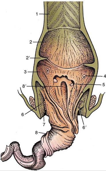
Figure 37-32 Cloaca of a drake with protruded phallus whose tip has been cut off, dorsal view. 1, Colon; 2, copro- deum; 2', coprourodeal fold; 3, urodeum; 4, ureteric orifice; 5, papilla of deferent duct; 6, proctodeum; 6', proctodeal glands; 7, lip of vent; 8, spiral groove of phallus; 8', beginning of spiral groove.
The gonad and tubular tracts of both male and female undergo remarkable involution outside the breeding season. These organs fill much of the body cavity while productive but, when inactive, regress to such an extent that they may be difficult to locate.
The Ovary
In the first 5 months after hatching, the ovary gradually develops from a small irregular structure with a finely granular surface to one in which individual follicles can be distinguished. These then rapidly increase in number and size until some are several centimeters in diameter (the size of an egg yolk; Figure 37-24/7 and Figures 37-33 and 37-34). The mature ovary resembles a truss of grapes, of various sizes, that is broadly attached to the cranial division of the left kidney. It contains several thousand follicles—far more than the number of eggs (about 1500) that even the most productive hen will lay. The larger follicles are pendulous and make contact with the stomach, spleen, and intestines. Each consists of a large, yolk-filled oocyte surrounded by a highly vascular follicular wall. Shortly before ovulation, a devascularized white band (stigma) appears opposite the stalk, indicating where the wall will rupture at ovula-
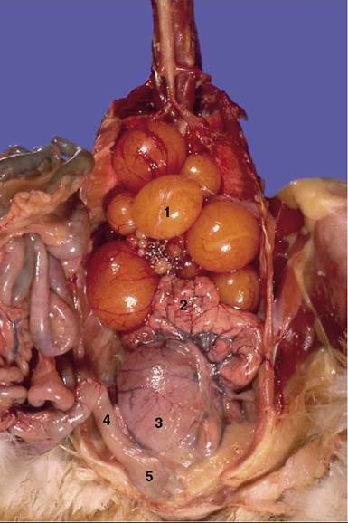
Figure 37-33 Ventral view of reproductive organs of a hen.
1, Ovary with follicles in different stages of development; 2, oviduct; 3, uterus; 4, colon; 5, cloaca.
tion (Figure 37-35/2 and Figure 37-34). The empty follicle (calix) regresses after ovulation and disappears in a few days. No corpus luteum is required because there is no embryo to maintain within the bird’s body.
The Oviduct
The oviduct is of much greater functional significance than its name implies. It not only conducts the fertilized ovum to the cloaca but also adds substantial amounts of nutrients (including the albumen); in addition, by enclosing the ovum with membranes and a shell, it provides protection for the developing embryo. It conveys spermatozoa to the ovum for immediate fertilization and may store them for a time for future use. (In the chicken, one insemination is sufficient to fertilize the ova released during the following 10 days or so.)
The oviduct (Figure 37-35/3-7 and Figures 37-33 and 37-34) may be divided into infundibulum, magnum, isthmus, uterus, and vagina according to the function of its parts; the uterus and vagina are, of course, not analogous to the like-named organs of mammals. The oviduct occupies the left dorsal part of the body cavity, where it is related to the kidney, intestines, and gizzard. It is a massive coil, approximately 60 cm long (i.e., about twice the body length) when fully functional but much
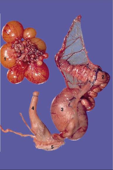
Figure 37-34 Isolated female reproductive organs. 1, Ovary with follicles in different stages of development; 2, oviduct; 3, uterus; 4, colon; 5, cloaca.
smaller in juveniles and during the nonlaying period. It is suspended from the roof of the body cavity by a peritoneal fold (mesoviductus), and some coils are connected by a continuation that forms the prominent muscular ventral ligament (Figure 37-35/12). The wall of the oviduct consists of the usual layers: serosa, tunica muscularis (consisting of outer spiral and inner circular layers), a scanty submucosa, and a tunica mucosa containing many glands.
The cranial end is formed by the 7-cm-long infundibulum (Figure 37-35/3), consisting of fluted and tubular parts.
The thin-walled fluted part is stretched to form a slit (infundibular ostium) several centimeters long; its lateral end is attached to the body wall near the last rib. The ostium is positioned by the left abdominal air sac in such a way that it can grasp newly released oocytes. The oocyte passes through the infundibulum in about 15 minutes. Fertilization must take place before the infundibular glands provide the chalaziferous layer, the thin coat of dense albumen directly around the yolk. (The chalazae, the coiled strands that suspend the yolk and allow it to rotate so that the germinal disk remains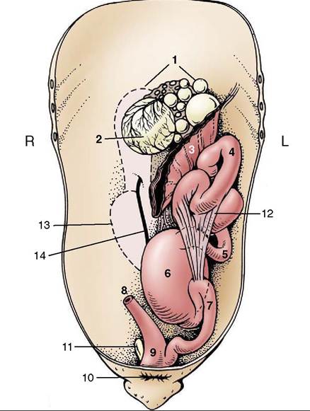
Figure 37-35 Ventral view of the reproductive organs of a laying hen, semischematic. 1, Ovary; 2, stigma on mature follicle; 3, infundibulum; 4, magnum; 5, isthmus; 6, uterus containing egg; 7, vagina; 8, colon; 9, cloaca; 10, vent; 11, vestigial right oviduct; 12, free border of ventral ligament of oviduct; 13, outline of right kidney; 14, right ureter.
uppermost, although part of this layer, develop farther along the genital tract (Figure 37-36Z3'). Some species have an infundibular sperm host gland in which sperm may be stored.
The highly coiled magnum (Figures 37-33, 37-34, and 37-35) measures about 30 cm and is the longest segment of the duct. Its walls carry massive mucosal folds and are thickened by the glands that contribute about half the total albumen to the egg. Calcium, sodium, and magnesium are also added here. The mucosal folds are lower and the secretion more mucous in the distal end of the magnum. The egg takes about 3 hours to pass through this part.
The isthmus (Figure 37-35Z5), about 8 cm long, is demarcated from the magnum by a narrow, translucent glandular zone. The isthmus, thinner and with lower mucosal folds than the magnum, secretes more albumen and also a material that rapidly congeals to form the two homogeneous membranes found between the albumen and the shell.
The egg takes upward of 1 hour to traverse the isthmus. The isthmus is lacking in psittacines.The isthmus is succeeded by the uterus (shell gland; Figure 37-35Z6), a thinner-walled, slightly enlarged chamber, about 8 cm long. Its mucosa bears many low folds and ridges that flatten themselves against the egg, which remains here for about 20 hours. Passing through the permeable membranes, some watery albumen is added to plump out the egg. This secretion is then followed by the deposition of the shell and shell pigments and an outer glazing or cuticle.
The final part, the vagina, (Figure 37-35Z7) is a muscular, S-shaped tube through which the completed egg
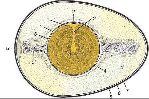
Figure 37-36 A semischematic section of a fertilized egg. 1, Yolk; 1', yolk membrane; 2, latebra; 2', germinal disk; 3, Chalaziferous layer; 3', chalaza; 4,4’, thin and dense albumen; 5, internal and external shell membranes; 5', air cell; 6, shell; 7, cuticle.
passes in seconds when it is laid. Its junction with the uterus is marked by a sphincter. Glandular crypts in the region of the sphincter have been found to store sperm. The vagina ends at a slitlike opening in the lateral wall of the urodeum. When the egg is laid (blunt end first), the vaginal opening protrudes through the vent, which minimizes contamination by the feces.
Sperm host glands may also be found at the uterovaginal junction where sperm can be stored for many months.
A remnant of the right oviduct (Figure 37-35Z11) is found on the right side of the cloaca; it may become cystic and enlarged.