INTRODUCTION
5.1.1 Lymphatic System
The lymphatic system is a complex network that helps maintain fluid balance, fights infections, and prevents diseases. It begins with the formation of interstitial fluid derived from capillary filtration and diffusion, resembling plasma but with lower protein content.
This fluid forms a gel-like matrix in the tissue spaces, which, under normal conditions, contains minimal free fluid. Lymphatic capillaries, the starting points of the lymphatic system, are fine vessels that arise from these interstitial spaces. They are equipped with one-way valves formed by overlapping endothelial cells, ensuring the unidirectional flow of lymph. Lymph capillaries collect about one-tenth of the fluid that leaks from the arterial end of blood capillaries, returning it to the bloodstream and bypassing the venous capillaries.Lymph flow is driven by low interstitial fluid pressure and can be significantly increased by factors such as muscle contractions, body movements, and changes in nearby arterial pressure. The composition of lymph is primarily water, with a small percentage of solids, including proteins, primarily albumin, and other molecules absorbed from the intercellular space. Lymph also contains white blood cells, particularly lymphocytes, which are crucial for the immune response.
Lymph nodes filter harmful substances and contain immune cells that fight infections throughout the body.
The glymphatic system, a component of the brain’s waste clearance pathway, is closely related to the lymphatic system. It plays a vital role in brain health by removing waste products from the brain into the cerebrospinal fluid.
The lymphatic system plays an important role in diseases, as it can be implicated in ailments such as lymphoma, which is a form of cancer that affects lymphocytes and organs within the lymphatic system.
It also plays a part in other diseases by maintaining fluid balance and supporting the immune system.5.1.2 Formation of Interstitial Fluid
The interstitial fluid obtained from capillary filtration and diffusion is similar to plasma but has lower protein concentrations due to capillary pores. The fluid is primarily located in the small spaces between proteoglycan filaments, forming a tissue gel. This tissue gel comprises a blend of proteoglycan filaments and trapped fluid. It possesses gel-like characteristics due to the high concentration of proteoglycan filaments that restrict the free flow of fluid. Due to thermal motion, the interstitial fluid generally moves through the tissue gel by diffusion, molecule by molecule. Though most of the fluid remains confined within the gel, small streams of free fluid and vesicles might be present. This free fluid can flow freely since it lacks proteoglycan molecules. In healthy animal tissues, free fluid is scarce, usually less than 1%. But, when tissues develop inflammation and edema, these small pockets and streams of free fluid level increase significantly. In such situations, it’s possible for as much as 50 percent or more of the fluid that causes edema to become free-flowing and separate from the proteoglycan filaments.
5.1.3 Lymphatic Capillaries
Lymphatic capillaries are the inception points of the lymphatic system, comprising a fine network of vessels that emerge from interstitial spaces. These capillaries are characterised by their blind-ended structures, termed capillary bulbs, which are fitted with valves that ensure lymph flows in only one direction. Muscle fibres encircle these bulbs, facilitating the propulsion of lymph through the vessels by contraction. The walls of lymphatic capillaries are formed by endothelial cells tethered to the surrounding connective tissue via anchoring filaments. These cells overlap at their edges, forming valves that permit the entry of interstitial fluid into the capillaries while preventing its exit, as backflow prompts the valves to seal shut.
Lymphatic capillaries merge to form progressively larger vessels by converging multiple tributaries. A fraction of the fluid that seeps out from the arterial end of blood capillaries, roughly one-tenth, is collected by these lymphatic capillaries and is subsequently returned to the bloodstream, bypassing the venous capillaries.
These capillaries, along with the larger lymph vessels, can actively pump lymph. When tissue swelling occurs due to an accumulation of fluid, the anchoring filaments exert a pull on the capillary walls, enabling fluid to enter through the endothelial junctions. Conversely, when tissue compression takes place, the internal pressure of the capillary increases, leading the endothelial cell edges to act as valves and push the lymph into the next segment of the lymphatic system, preventing retrograde flow. Additionally, lymphatic capillary endothelial cells contain contractile actomyosin filaments, which are thought to contribute to the rhythmic contractions that facilitate lymph movement, akin to the contractions observed in smaller blood vessels and larger lymphatic vessels.
38
DOI: 10.1201/9781003426851-5
Particular body structures, including the outermost layers of the skin, the central nervous system, the cornea, bones, and the alveoli of lungs, lack lymphatic vessels.
5.1.4 Lymph Flow
The lymphatic system functions under significantly low interstitial fluid pressure. The pressure inside the capillaries is usually around 1 mm HG during resting conditions. However, lymph flow can increase by more than twenty times when this pressure rises to atmospheric levels. This increase in lymph flow is facilitated by various factors such as elevated capillary hydrostatic pressure, increased plasma colloid osmotic pressure, increased interstitial fluid colloid osmotic pressure, and increased capillary permeability. All these factors contribute to an increase in interstitial fluid volume and pressure.
As lymph vessel segments fill with fluid, they stretch.
This stretching triggers the smooth muscle in the vessel wall to contract, pushing the fluid through the next valve into the following lymphatic segment. This process repeats along the lymph vessels until the fluid drains into the bloodstream.There is a considerable variation in the flow of lymph in different animals. This variation depends on the animal’s age, size, and lactation stage. For instance, in cows, lymph flow from the udder ranges from 13 to 45 ml/h and is characterised by pulsatile episodes with monophasic and multiphasic patterns. In calves, when fed milk, lymph flow from the thoracic or intestinal ducts averages 695 ml/hr and 429 ml/hr, respectively. However, this flow increases to 899 ml/hr and 517 ml/hr when they are fed hay and grain. Interestingly, the lymph flow increases after whole milk feeding, but there is no consistent diurnal pattern in lymph flow associated with feed intake in calves fed hay and grain. In dogs, the volume of lymph in a dog’s kidney is about 1% of the blood volume in the cortical peritubular capillaries. Additionally, adult dogs have three times lower lymph flow compared to puppies.
The lymphatic system plays a crucial role in transporting large molecules like proteins that tissues, in any other way, cannot absorb. Lymphatic flow is facilitated by the natural contractions of the lymph vessels and external pressures that intermittently compress these vessels. The following are the external factors that contribute to this process in order of significance: muscle contractions of the muscles surrounding the lymph vessels, body movements, alteration of pressure in nearby arteries, and external pressure applied to the body. During physical activity, the lymphatic system’s pumping action is greatly enhanced, potentially increasing lymph flow by 10 to 30 times. In contrast, the movement of lymph tends to be much slower when the body is at rest.
5.1.5 Composition of Lymph
Lymph is a transparent and colourless fluid consisting of 96% water and 4% solids.
At its most basic level, lymph is just interstitial fluid that contains an additional 20g/L of protein, primarily albumin. Due to the large size of its molecules, not much fibrinogen can enter the lymphatics. However, even with minimal fibrinogen and without any platelets, lymph can still clot under certain conditions. The average protein concentration in lymph is 4-5 g/100 ml in the thoracic and intestinal ducts, regardless of whether the diet is milk-fed or grain-fed.Additionally, lymph accumulates other proteins in the intercellular space. These are not like the plasma proteins, which are intentionally secreted into the bloodstream for some purpose. These are products of lytic extracellular matrix processing, tissue remodeling, cellular metabolic activities and the products of cell death. The vast majority of these proteins will undergo phagocytosis by tissue macrophages and antigen-presenting cells long before they reach the lymph nodes, let alone the venous circulation, which is why they never appear in the blood. The proteins of lymph have a lot of regional variation.
Lipid in lymph averaged 0.79 g- 3.14/100 ml in the thoracic and intestinal ducts. Calves that were fed milk consumed 2 to 3 times more lipid compared to those that were fed hay and grain. However, there was not much variation in lipid output throughout the day in the lymph of calves fed either diet. Lymph is modified by passage through the lymph node, whereas data concerning possible changes in the collecting lymphatics are less conclusive.
5.1.6 Lymph Nodes
Lymph nodes, also known as lymphatic nodes or lymph glands, are small glandular structures that are a part of the lymphatic system. A dense connective tissue capsule encloses them and consists of three layers: the cortex, paracortex, and medulla (Figure 5.1).
The cortex of a lymph node comprises primary and secondary lymphoid follicles. Initially, the primary follicle takes shape; later, when antigens infiltrate the body and make their way to the lymph nodes, the cells in the germinal centre region of the primary follicle begin to proliferate.
After this multiplication, the primary follicles become secondary follicles. The cortex also contains B lymphocytes, typically grouped into the primary follicles and macrophages.The paracortex between the cortex and medulla, is rich in T lymphocytes. The medulla, the innermost layer of the
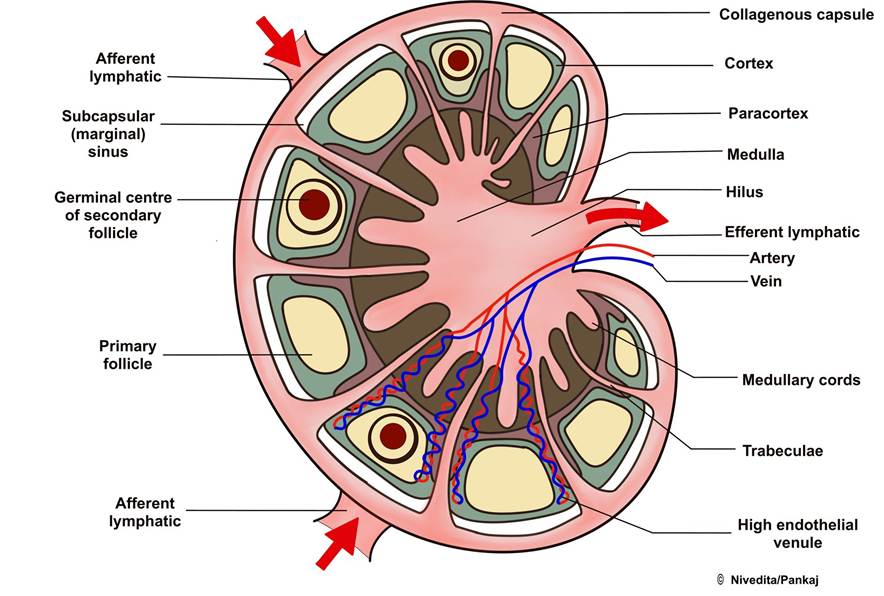
FIGURE 5.1 The structure of the lymph node is arranged in layers of capsule, cortex and medulla
lymph node, contains B and T lymphocytes, macrophages, and large blood vessels. The blood vessels of the lymph node pass through the medulla.
Lymph nodes get lymph through one or two afferent vessels, which divide into small channels. The lymph moves through these vessels and channels, reaching the cortex, and then circulates through the lymph node’s cortex, paracortex, and medulla. From the medulla, the lymph exits the node through one or two efferent vessels.
Lymph nodes act as filters, with their primary role being to eliminate bacteria and harmful substances from the lymphatic fluid. As lymph travels through the lymph nodes, it undergoes a filtration process where water and electrolytes are separated, and proteins and lipids are kept within the lymph. Bacteria and other toxic substances are destroyed by the macrophages of the lymph nodes, making them important defence barriers. During an infection or other processes in a specific body region, the lymph nodes’ activities in that region increase, causing the lymph nodes to swell.
5.1.7 Role of the Glymphatic System in the Brain
Iliff and colleagues have coined the term glymphatic system, which refers to a perivascular pathway that spans the whole brain and has a vital function in removing interstitial solutes and waste materials from the brain and transporting them into the cerebrospinal fluid (CSF). This system is facilitated by aquaporin-4 (AQP4) channels, which are densely expressed in astrocyte end-foot processes. The deletion of these water channels was found to impair the clearance of extracellular solutes, including amyloid-β (Aβ), a protein associated with Alzheimer’s disease.
The glymphatic system serves multiple purposes in neurophysiology. It is essential for delivering nutrients, mainly glucose, the circulation and distribution of apolipoprotein E isoforms produced by the choroid plexus, and astrocytic paracrine signalling with lipid molecules. However, its fundamental role is clearing extracellular metabolites and waste products from the brain parenchyma into the CSF.
The glymphatic system’s function is crucial during sleep, where it aids in the clearance of interstitial lactate that accumulates during regular daily brain activity. Disruptions to the glymphatic pathway, such as AQP4 knockout, CSF volume reduction, or alteration of posture, can lead to increased brain lactate levels and reduced cervical lymph node lactate levels.
Impairment of the glymphatic system has been linked to various central nervous system diseases. The influx of glymphatic CSF is reduced in the brain of elderly individuals and in a transgenic mouse model of Alzheimer’s disease. As a result, the clearance of Aβ is significantly impaired in both cases. Furthermore, glymphatic impairment is a prominent feature of cerebrovascular disease, including both subarachnoid haemorrhage and multiple microinfarctions. After a traumatic brain injury, reduced clearance of interstitial solutes in the cortex was linked to neurological deficits in both motor and cognitive function.
5.1.7 Lymphatic System Disease
Lymphoma encompasses a group of cancers that arise from the abnormal growth of lymphocytes. Lymphocytes affect organs such as the spleen, bone marrow, and lymph nodes. While most lymphoma cancers occur in these organs, any part of the body can be affected by the disease. Multicentric lymphoma, Alimentary lymphoma, Mediastinal lymphoma, and Extranodal lymphoma are the four most prevalent types of lymphoma in dogs.
Mammary tumours can change the way lymphatic fluid flows, leading to the creation of new drainage paths and the involvement of a large number of lymph nodes. Therefore, understanding the lymphatic drainage of the affected mammary glands is crucial for the surgeon to perform the appropriate surgery and determine the prognosis after the operation. Laverdia-CA1 (Verdinexor tablets) is a medication used to treat lymphoma, which is a type of cancer affecting the lymph nodes and lymphatic system in dogs.
The lymphatic system is essential in maintaining the balance of interstitial fluid protein concentration, volume, and pressure. It acts as a safety valve, returning excess proteins and fluids from the tissue spaces back to the bloodstream, thereby controlling protein concentration. Additionally, it helps absorb fats and fat-soluble vitamins from the digestive system. It supports the immune response by producing lymphocytes and antibodies to combat harmful substances in the interstitial fluid, volume, and pressure.
5.2
More on the topic INTRODUCTION:
- Introduction
- Introduction
- EPIDEMIOLOGY
- The Doe
- Vogelnest L., Portas T. (Eds.). Current Therapy in Medicine of Australian Mammals. CSIRO,2025. — 848 p., 2025
- NON-INFECTIOUS DISEASE
- Parenteral Nutrition in Ruminants
- Diseases Caused by Bovine Viral Diarrhea Virus (BVDV)
- Smith Bradford P., Van Metre David C., Pusterla Nicola (eds.). Large Animal Internal Medicine. Part 2. 6th edition. — Elsevier,2020. — 2279 p., 2020
- Other Problems Affecting the Buck