REGULATION OF RENAL BLOOD FLOW
Renal blood flow is regulated mainly by autoregulation. Autoregulation is the intrinsic ability of an organ to regulate its own blood flow. Some vital organs in the body, such as brain, heart, and kidneys, have autoregulation mechanisms.
It is highly significant and more efficient in the kidneys. Renal autoregulation is important in maintaining the glomerular filtration rate (GFR). Blood flow to the kidneys remains normal even when the mean arterial blood pressure varies widely. This helps maintain normal GFR.Two mechanisms are involved in renal autoregulation: Myogenic response: In the afferent arteriole, increased
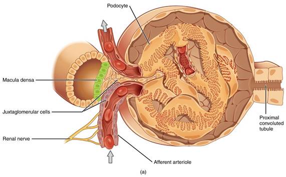
blood pressure would increase the stretch, and the arteriole would respond by contracting. In this way, renal blood flow would be decreased. A reduction in blood pressure would cause less tension, and the blood vessel would dilate, hence increasing renal blood flow. Tubuloglomerular feedback: The macula densa plays an important role in tubuloglo- merular feedback, which controls the renal blood flow and GFR. Macula densa cells sense changes in volume delivery as well as NaCl concentration to the distal tubules.
18.13.1 Urine Formation
Urine formation consists of three processes: glomerular filtration, tubular reabsorption from the renal tubules into the blood, and tubular secretion from the blood into the renal tubules. Filtration is the function of the glomerulus, whereas reabsorption and secretion are the functions of the tubular portion of the nephron.
18.13.2 Glomerular Filtration
Most substances must be cleared from the blood, especially the end products of metabolism such as urea, creatinine, uric acid, and urates. These substances are poorly reabsorbed and therefore excreted in large amounts in the urine.
Certain foreign substances and drugs are also poorly reabsorbed, so their excretion rates are high. Electrolytes like sodium ions, chloride ions, and bicarbonate ions are highly reabsorbed from renal tubule, so that only very slight amounts appear in the urine. Substances such as amino acids and glucose are completely reabsorbed from the renal tubules and hence do not appear in the urine.The formation of urine begins when an ultrafiltrate of plasma passes through the fenestrated capillary endothelium, the basement membrane, and the epithelium of Bowman’s capsule. The glomerular capillaries are impermeable to proteins, so the glomerular filtrate is protein-free and devoid of cellular elements, including RBCs. Other constituents of the glomerular filtrate are similar to the concentrations in the blood plasma
Size of solute is an important parameter for their filtration. Filtration of solutes is inversely proportional to their size. The barrier present in glomerular membrane is selective in determining the molecules to filter, based on their size and electrical charge. Negatively charged large molecules are filtered less easily than positively charged molecules of equal molecular size. Albumin is restricted from filtration, because of its negative charge and the size. In certain kidney diseases, the negative charges on the basement membrane are lost and as a result of this loss, some of the lower-molecular-weight proteins, especially albumin, are filtered and appear in the urine.
18.14
More on the topic REGULATION OF RENAL BLOOD FLOW:
- REGULATION OF RENAL BLOOD FLOW
- THE JUXTAGLOMERULAR APPARATUS
- Urine Formation
- Regulation of Arterial Blood Pressure and Blood Volume
- Rana Tanmoy (ed.). Principles of Veterinary Animal Physiology. CRC Press,2026. — 290 p., 2026
- Solute Reabsorption by the Medullary ThickAscending Limb and Collecting Duct Generates and Maintains Medullary Hypertonicity
- Functional Morphology of Kidney
- GLOMERULAR FILTRATION
- SELF EVALUATION - CHAPTER 11
- Avian Renal Physiology