MILK SYNTHESIS AND SECRETION AND ITS COMPOSITION
22.15.1 Secretion and its Constituents
Milk secretion is the process in which the milk constituents synthesized by epithelial cells are released into the alveoli’s lumen. Most of these constituents originate from the alveolus itself.
While milk fat, protein, and lactose are produced within the alveolar epithelial cells, certain elements like vitamins, minerals, and specific proteins are not manufactured in the mammary gland but are instead sourced from the bloodstream. These non-synthesized components enter the cells from the blood and are then discharged into the milk. Lactose, fat, and the majority of proteins are created in the epithelial cells using precursors supplied by the bloodstream.The secretion of milk constituents, including fat and protein, involves distinct processes within the epithelial cells. Milk fat forms as small droplets within these cells, which then merge as they move towards the cell’s apical part. These fat droplets are subsequently released from the epithelial cell through a pinching-off mechanism (Figure 13). During this process, the fine apical membrane of the epithelial cell encases the milk fat globule’s outer layer.
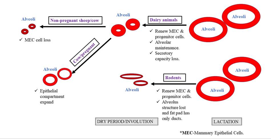
FIGURE 22.6 Diagram showing involution of the mammary gland in bovine and rodents.
Casein and lactose molecules are released as secretory vesicles surrounded by a membrane. These vesicles migrate towards the apical part of the cell before being discharged into the alveolar lumen. Before release, the secretory vesicle’s wall fuses with the cell membrane, and its contents are discharged into the alveolar cavity, repairing the membrane loss caused by the release of fat droplets. This secretion process is continuous, with each epithelial cell contributing to the overall secretion.
The molecules of proteins, milk sugar, vitamins, and minerals are evenly distributed within the milk.
However, milk fat globules float in water upon entering the alveolar lumen. These fat droplets aggregate into small clusters that cannot easily pass through the ducts draining the alveoli. Meanwhile, water flows freely along the ducts, gradually filling the cisterns of quarters. Some fat remains within the secretory cell and is only released when udder pressure decreases during milking. Consequently, the milk initially drawn from the udder contains only 1 to 2% fat, with subsequent milk possibly containing more fat.22.15.2 Rate of Secretion of Milk
Epithelial cells display a heightened milk synthesis rate within the first 10 hours post-milking, which gradually diminishes afterward. When a cow or buffalo goes unmilked, milk synthesis and secretion typically cease within a specific timeframe following the last milking session. The cessation of milk secretion is due to increased pressure within the alveolus, which constricts blood vessels, hindering the flow of blood and the supply of milk precursors to epithelial cells. Low-producing cows are less affected by extended milk intervals due to having less secretory tissue in the udder. Consequently, younger and low-producing cows experience a greater increase in udder pressure per unit milk, underscoring the importance of short milking intervals for such animals.
Leaving a significant amount of milk (2kg or more) in the udder after incomplete milking can lead to a permanent reduction in milk production. Milk secretion is influenced by various factors including parity, lactation stage, milking interval, frequency of milking, residual milk, and milking management practices. The rate of milk secretion plays a crucial role in determining the additional milk yield achievable with more frequent milking. For instance, milking high-producing cows (>12-15kg∕day) three times daily instead of twice daily can result in a 20% increase in milk yield. Further increases in milk yield (5 to 10%) can be achieved by milking four times per day.
However, considering feeding and labor costs, milking four times per day may not be economically feasible or advisable. Economically, three times daily milking becomes favorable for cows producing over 15kg∕day. Sleeping one milking session per week may lead to a reduction in total milk yield by 5 to 10%. Maintaining regular milking intervals of 12 hours for cows and buffaloes with moderate production potential is crucial in dairy farming, particularly in rural settings. Agalactia refers to complete lactation failure, while hypogalactia denotes partial lactation failure.22.15.3 Role of Nerves in Milk
Secretion Regulation
The regulation of milk secretion by the nervous system has not been elucidated extensively. Many studies indicate that frequent milking of dairy animals stimulates milk secretion. Mammary glands respond to the following neural factors for milk synthesis.
It has been observed that the production capacity of the mammary gland in rats can be modulated by the nutritional demand of the pups or litter. In ruminants, increased milking frequency (IMF) of 2-3 times or intense suckling by the calf results in increased milk synthesis. Thus, IMF can be utilized to augment milk production and production efficiency.
In dairy animals, such as cows, milking three times a day results in 15-20% more milk production. Similarly, milking four times is preferable to three times, leading to an additional increase in milk production. This indicates that the mammary gland is especially sensitive to the demand of offspring in early lactation, influencing the shape of the lactation curve and lactation persistency.
Increased milking frequency (IMF) in ruminants and rodents leads to:
1.An increase in mammary cell number
2. An increase in mammary cell activity
3. Additionally, the effect of suckling intensity on mammary gland development has also been reported.
In rodents, mammary development with increased suckling is accompanied by:
1.An increase in DNA
2. An increase in RNA
3. An increase in the RNA:DNA ratio
22.15.4 Regarding the Impact of IMF and
Suckling Intensity on Hormones
In rodents, the act of suckling prompts the release of both prolactin and oxytocin, wherein prolactin serves to stimulate mammary cell differentiation. An increased litter size is associated with elevated prolactin levels within the pituitary gland. This elevation in prolactin levels is linked to increased mammary cell activity. When oxytocin is injected into rats, it prolongs lactation by facilitating the complete evacuation of the mammary gland, although it does not impact milk yield. During late lactation in rodents, cortisol is recognized as the limiting factor; however, administering cortisol does not consistently result in a significant increase in milk yield. This suggests that local factors play a crucial role in regulating the mammary response to systemic hormones.
22.16
More on the topic MILK SYNTHESIS AND SECRETION AND ITS COMPOSITION:
- Anatomic Structures of the Bovine Mammary Gland
- Protein
- Diagnosis of Mastitis
- References
- Trace Minerals
- Dairy Goat Herd Health Management and Preventive Medicine
- Non-mastitic Alterations in Goat Milk
- Abnormalities of the Ovaries
- Winter Dysentery in Cattle (Bovine Coronavirus)
- Fluid and Electrolyte Balance